Findings
CT
- Ill-defined hyperattenuating mass in the superior aspect of the right cerebellar hemisphere
- Surrounding parenchymal edema
- Corresponding mass effect with crowding of the fourth ventricle and displacement of the cerebellar tonsils 13 mm below the foramen magnum
- Likely mild corresponding obstructive hydrocephalus
MRI
- Ill-defined T2/FLAIR hyperintense mass in the superior aspect of the right cerebellar hemisphere and superior aspect of the vermis measuring approximately 4.3 x 2.2 x 4.2 cm
- Nodular internal areas of restricted diffusion and corresponding patchy enhancement
- Peripheral susceptibility artifact about one of these nodular areas in the anterior aspect of the right cerebellar hemisphere, likely relating to hemorrhage
- Thickening and enhancement of the adjacent tentorial leaflet
- FLAIR signal hyperintensity in a right occipital sulcus and possible adjacent cortical FLAIR signal hyperintensity without definite corresponding restricted diffusion or enhancement (possibly representing leptomeningeal spread)
- Small focus of restricted diffusion along the falx without definite corresponding enhancement (also possibly a leptomeningeal tumor deposit)
- Associated mass effect in the posterior fossa resulting in cerebellar tonsillar herniation and crowding of the fourth ventricle with mild obstructive hydrocephalus
Annotated Images & Illustrations
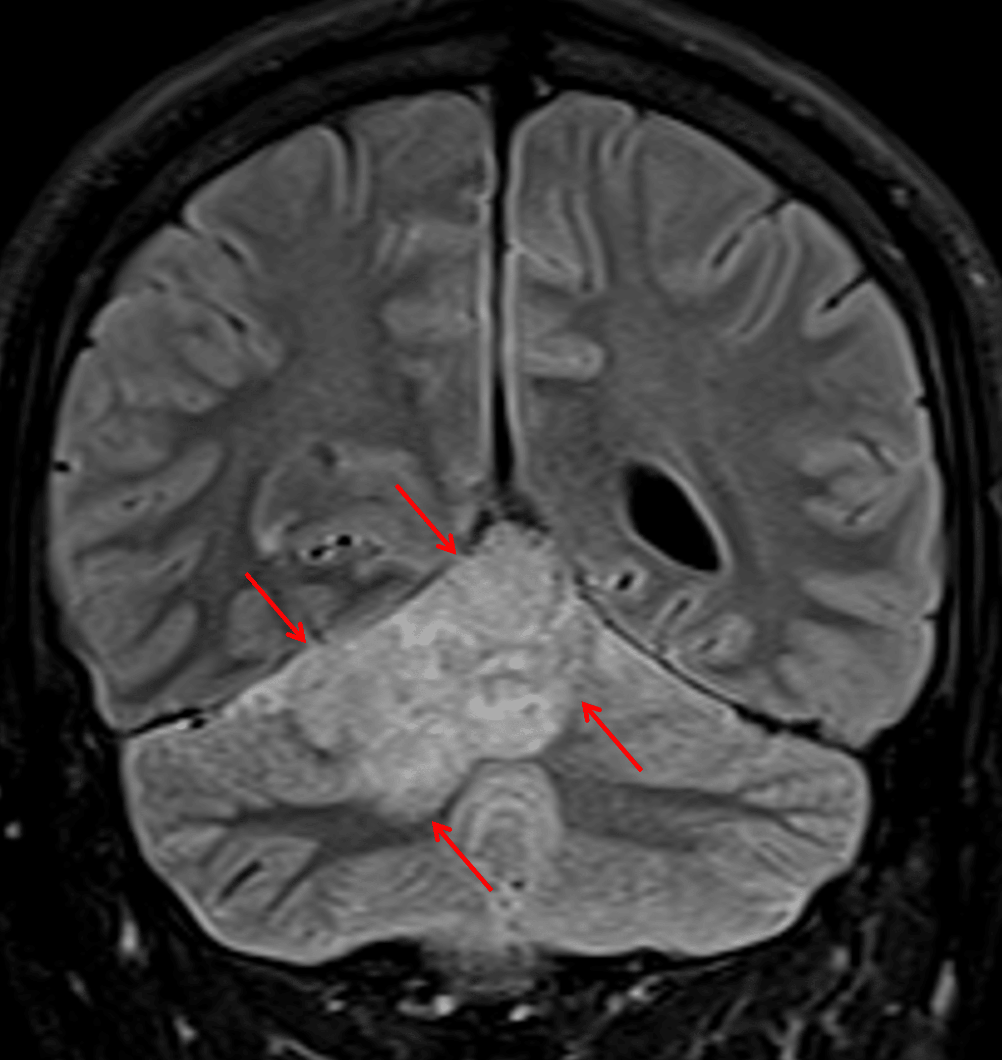
T2/FLAIR hyperintense mass in the superior aspect of the right cerebellar hemisphere and vermis (red arrows).
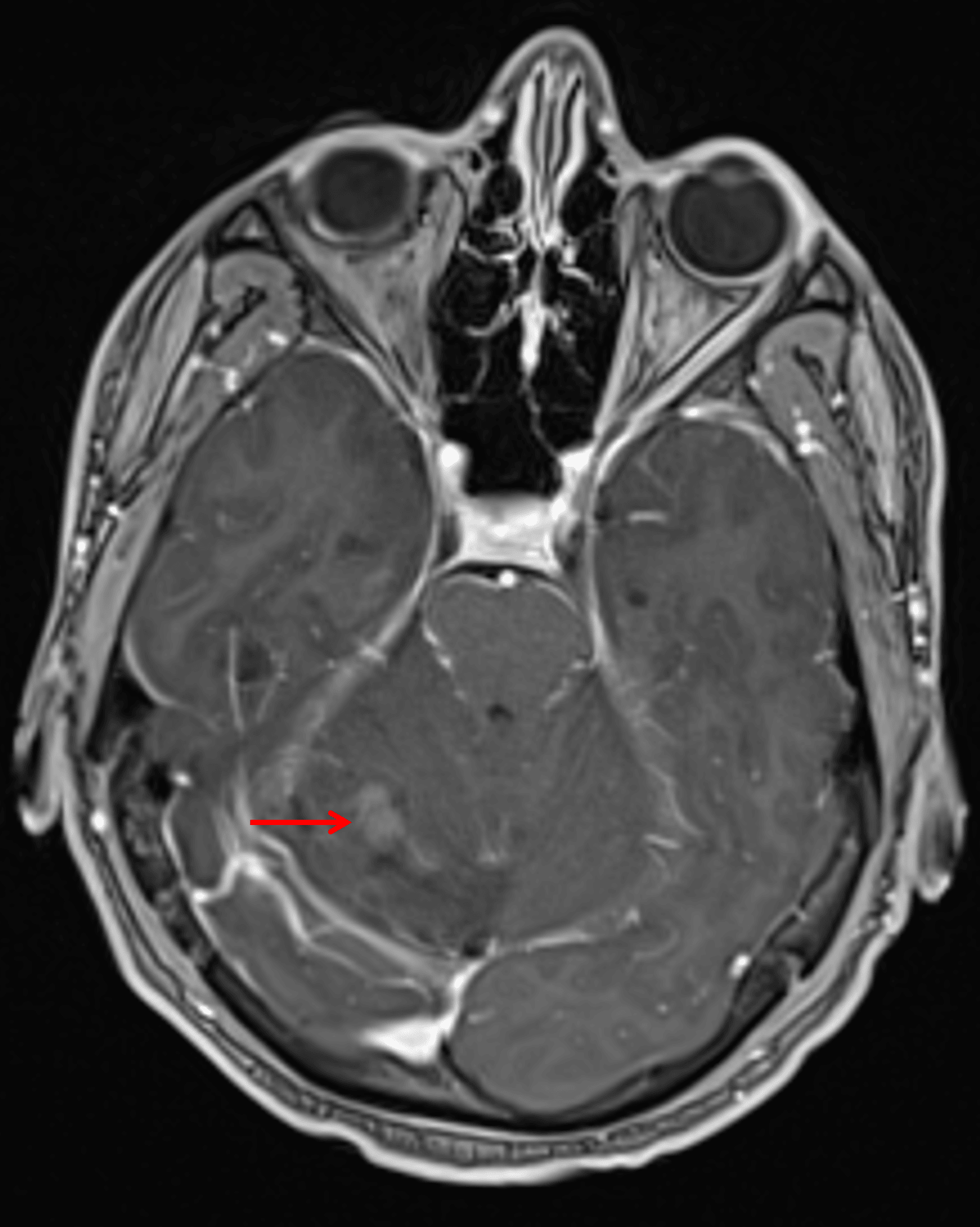
Mild corresponding patchy enhancement (red arrow).
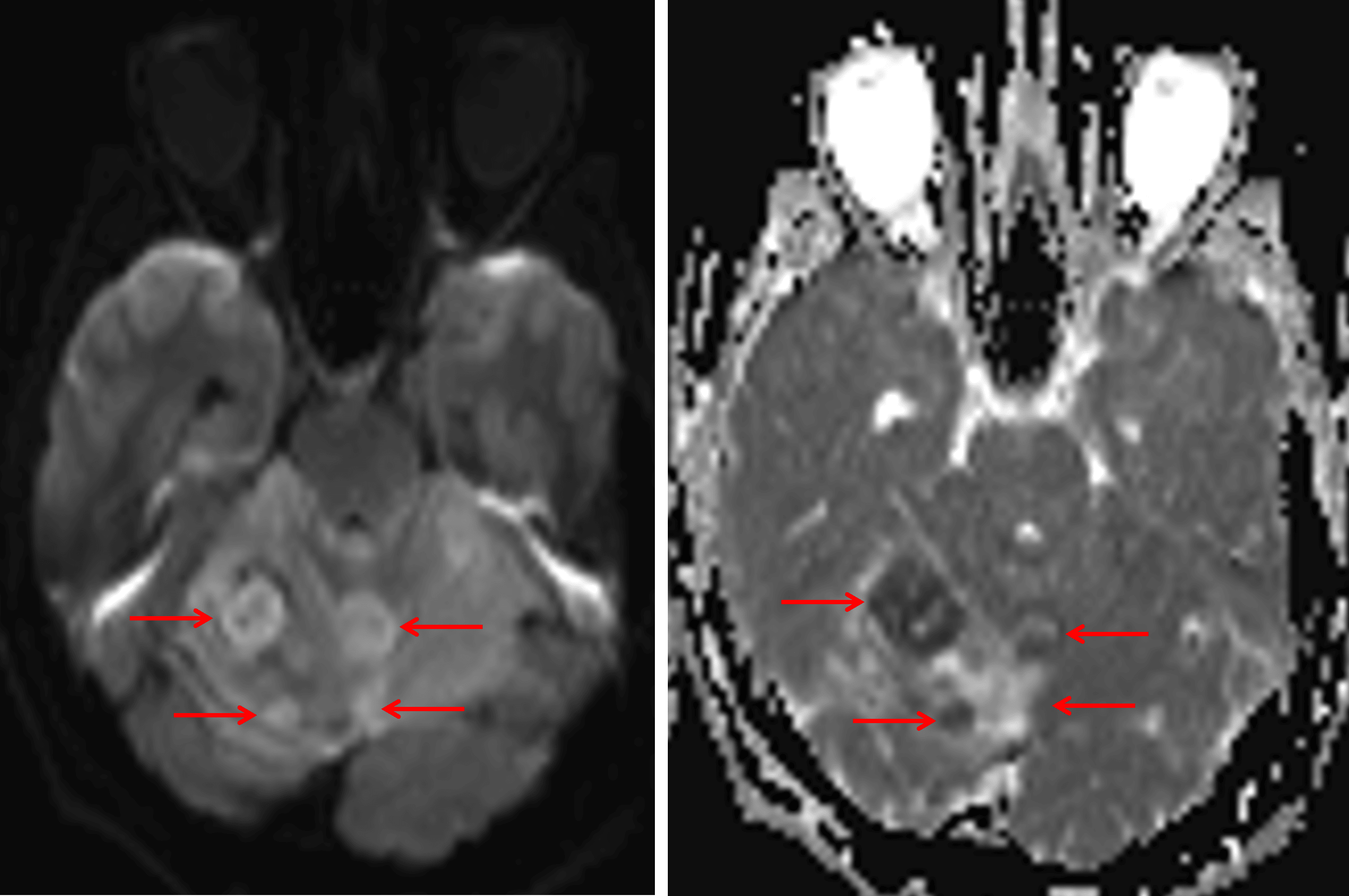
Internal nodular areas of restricted diffusion (red arrows).
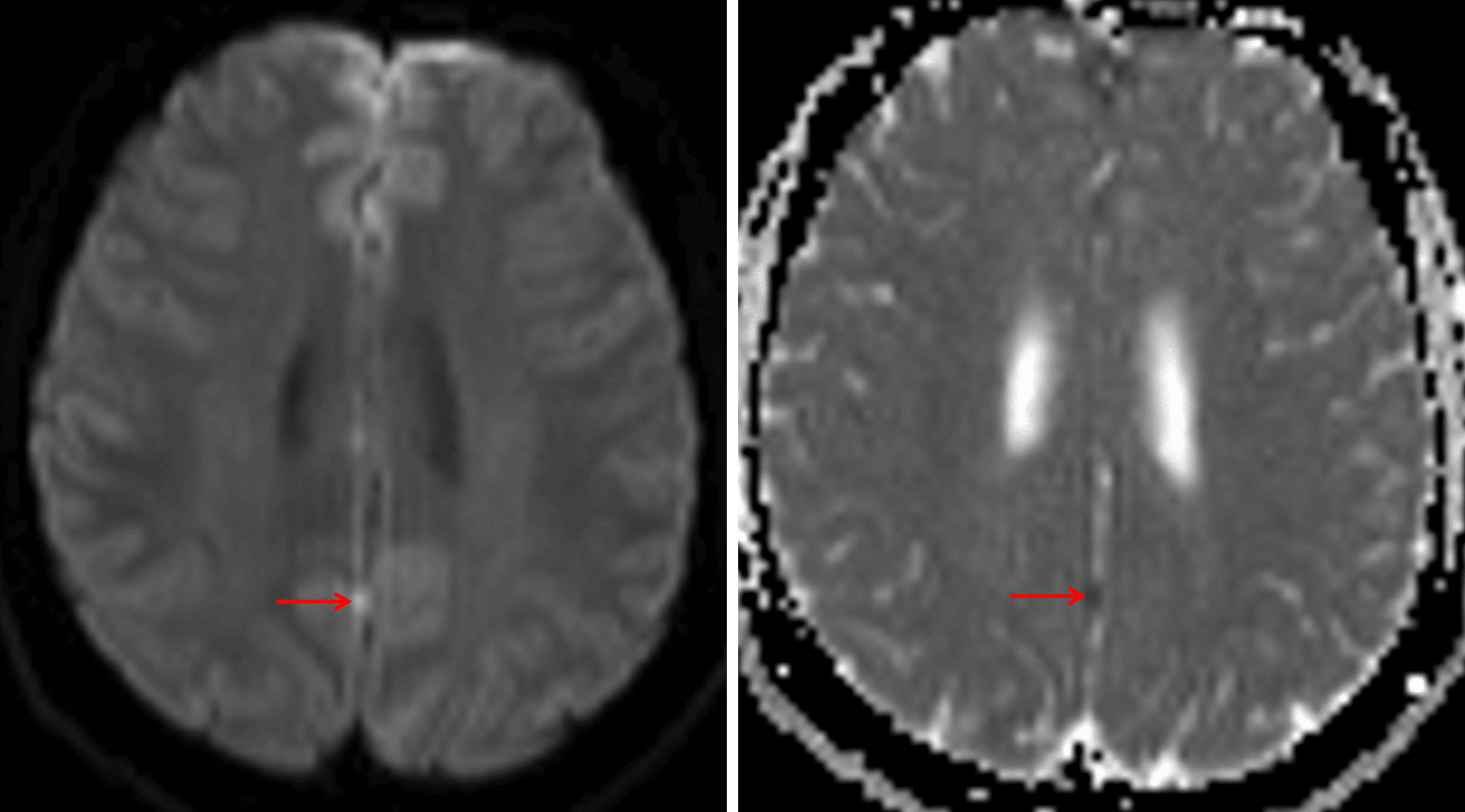
Nodular focus of restricted diffusion along the interhemispheric fissure (red arrows), which is concerning for a leptomeningeal tumor deposit.
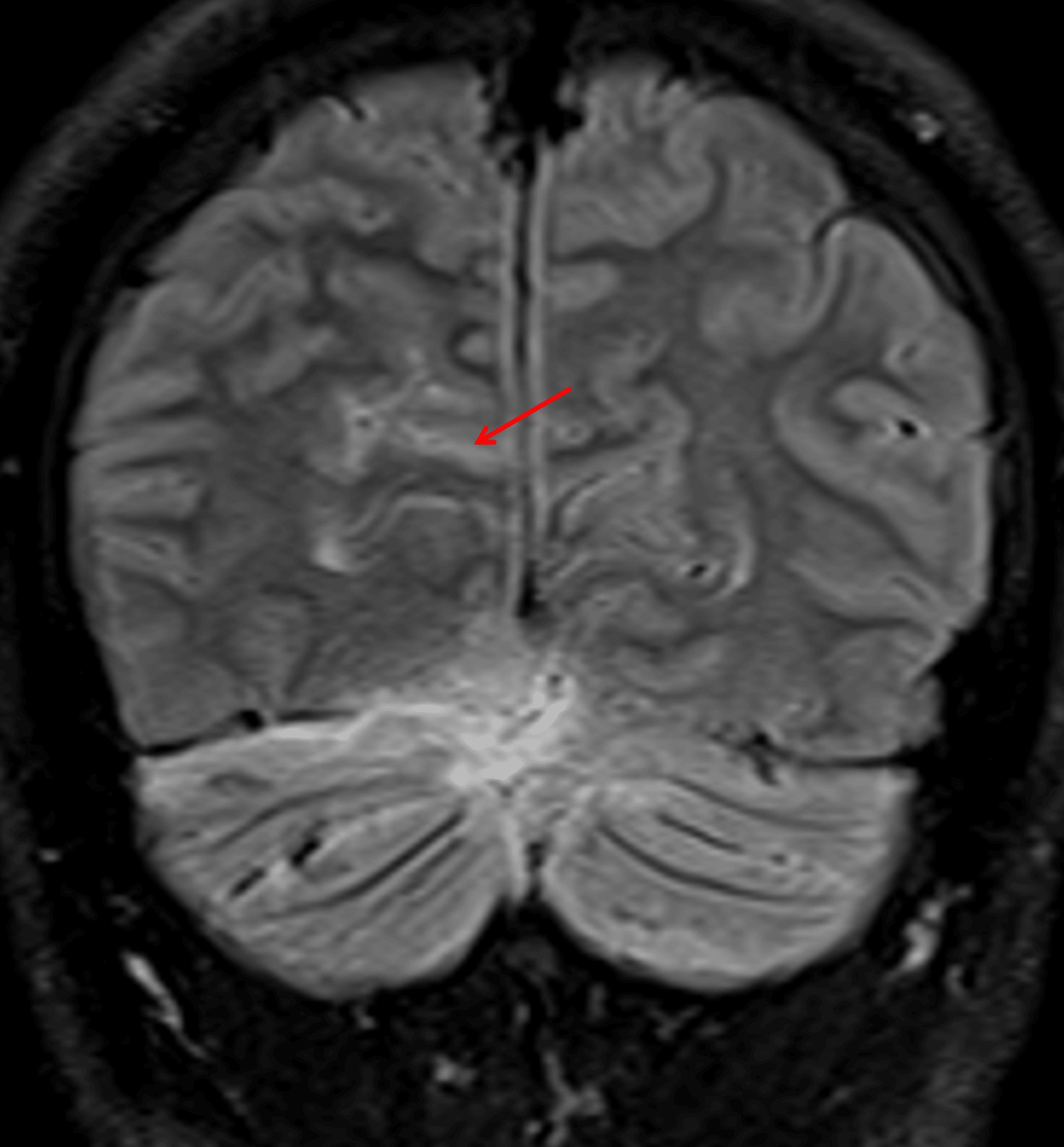
FLAIR signal hyperintensity in a right occipital sulcus (red arrow), which may also relate to leptomeningeal tumor dissemination.
Diagnosis
Medulloblastoma - most likely SHH-activated (TP53 wild-type) or Group 4
Differential Diagnosis
Become a PRO member to unlock the differential diagnosis
Pearls
Become a PRO member to unlock the pearls