Demographics:
11 years old, Male
Indication:
Diabetes insipidus
Findings
- Thickening of the pituitary stalk with corresponding diffuse enhancement
- Absent posterior pituitary bright spot on pre-contrast T1-weighted imaging
- Confluent T2/FLAIR signal hyperintensity in the bilateral cerebral periventricular white matter with white matter volume loss and thinning of the corpus callosum (patient had a known history of periventricular leukomalacia)
- Few scattered foci of susceptibility artifact in the left greater than right parietal lobes
Annotated Images & Illustrations
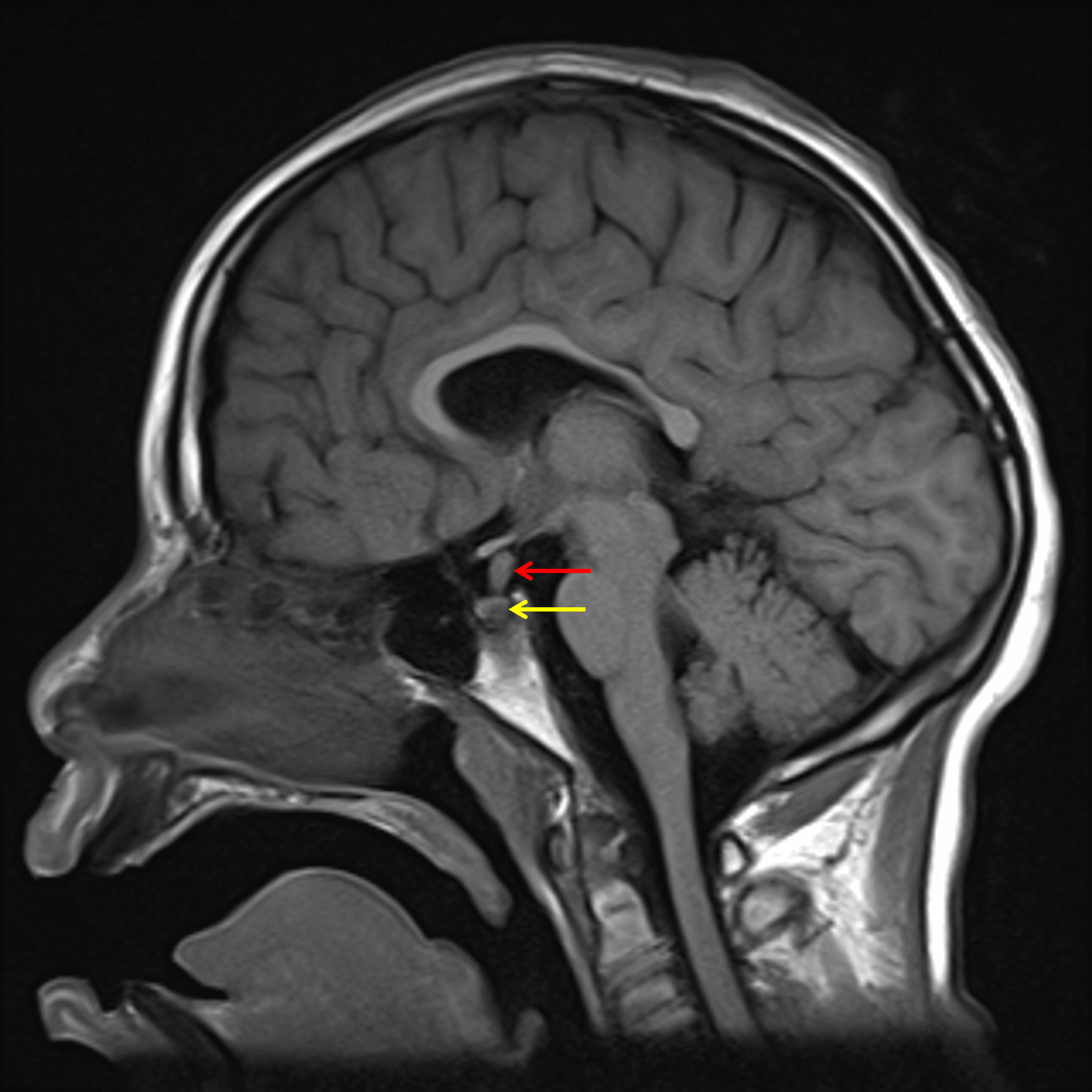
Thickening of the pituitary stalk (red arrow) with absent posterior pituitary T1 bright spot (yellow arrow).
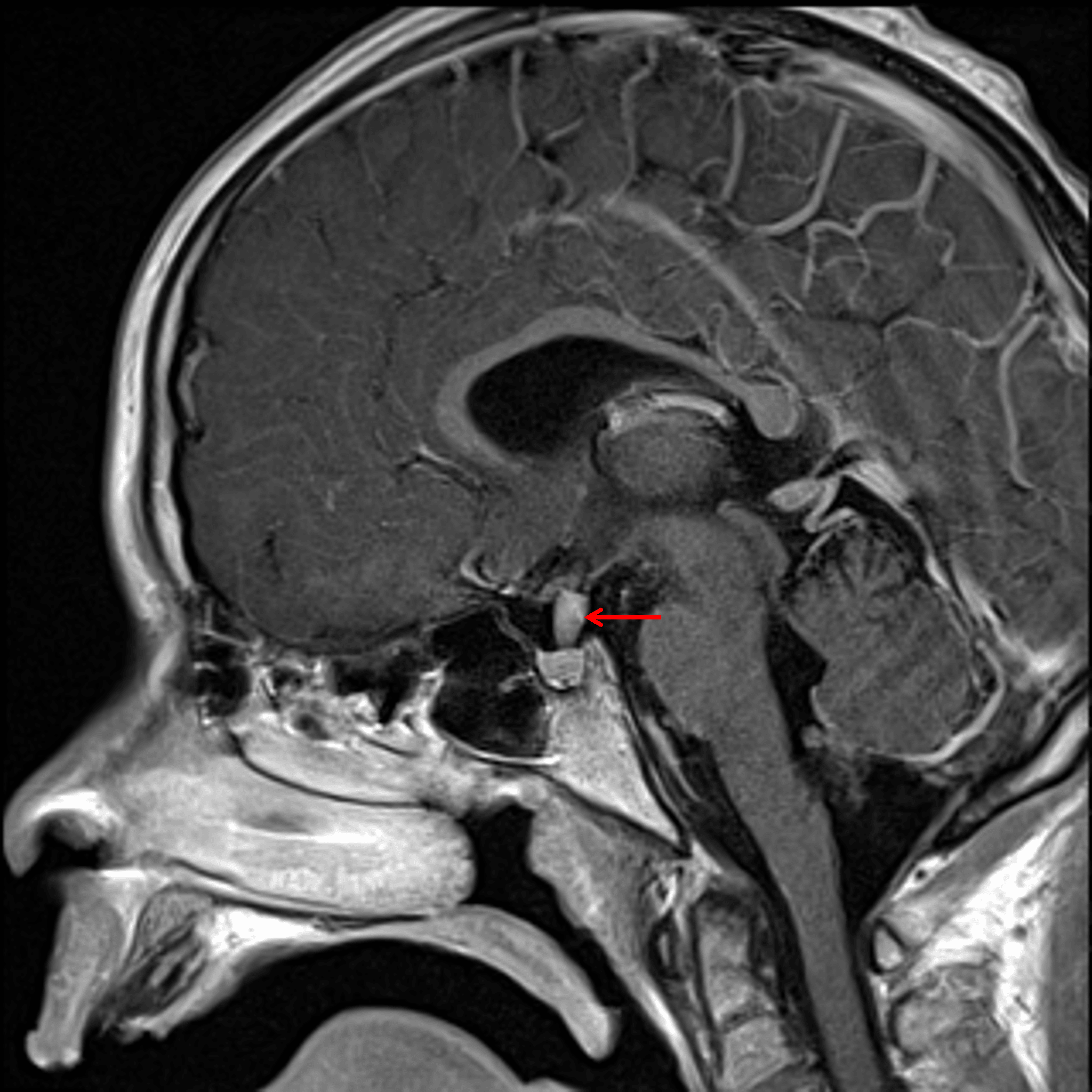
Diffuse enhancement of the pituitary stalk lesion (red arrow).
Diagnosis
Langerhans cell histiocytosis (LCH)
Key Imaging Features
Become a PRO member to unlock the key imaging features
Differential Diagnosis
Become a PRO member to unlock the differential diagnosis
Discussion
Pearls
Become a PRO member to unlock the pearls