Demographics:
13 years old, Male
Indication:
Headache, history of renal transplant
Findings
CT
- Numerous hyperattenuating foci in the cerebral and cerebellar white matter bilaterally with associated vasogenic edema
MRI
- Numerous enhancing lesions in the bilateral cerebral and cerebellar hemispheres, the cerebellar vermis, and the medulla, many of which are periventricular in location and extend to the ependymal surface of the ventricles
- The majority of these lesions demonstrate solid enhancement, though a few demonstrate peripheral enhancement
- Variable corresponding restricted diffusion
- Corresponding vasogenic edema and mass effect without midline shift or evidence of herniation or hydrocephalus
Annotated Images & Illustrations
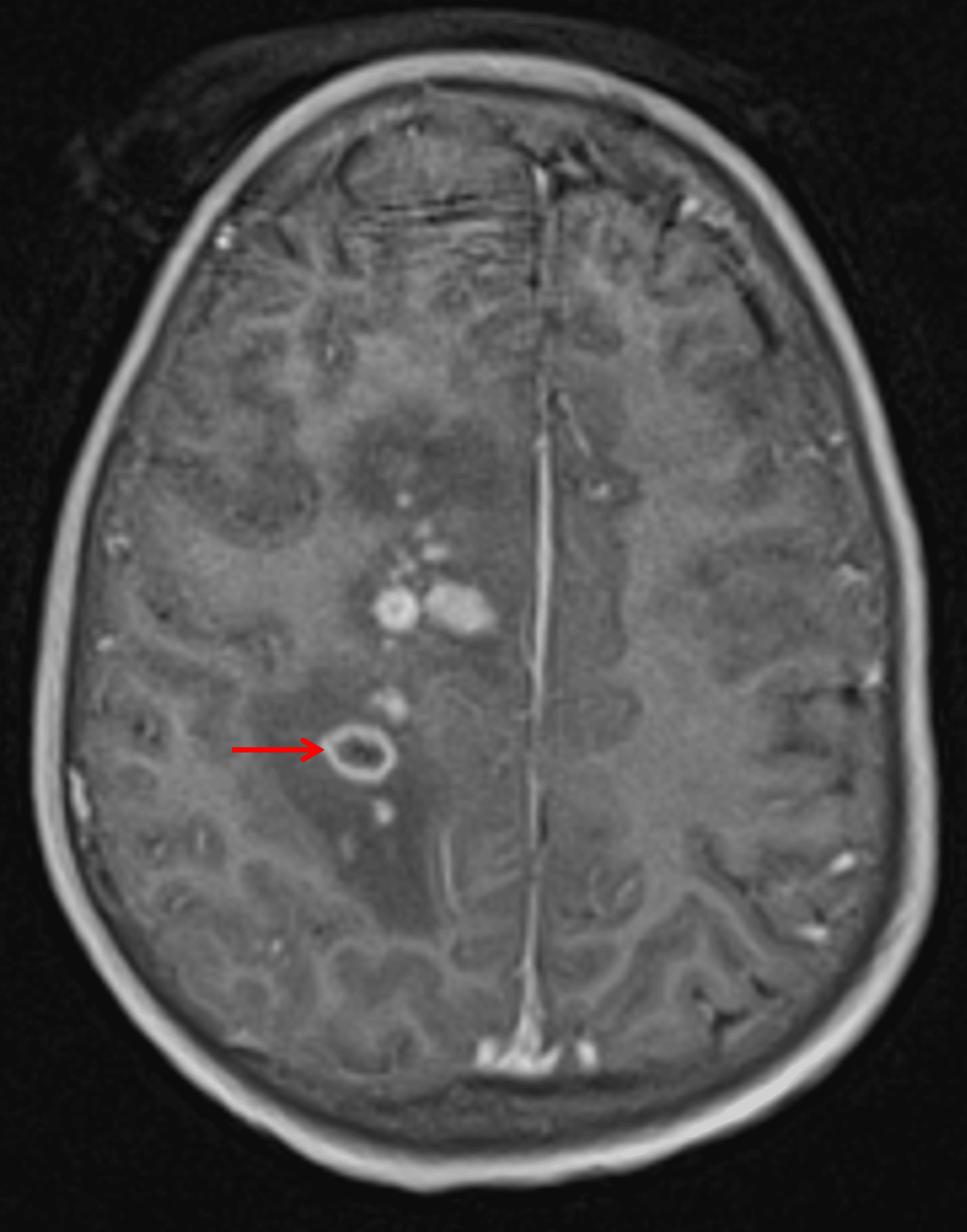
Numerous small enhancing lesions are seen. Most solidly enhance, but one in this image demonstrates peripheral enhancement (red arrow).
Diagnosis
Post-transplant lymphoproliferative disorder (PTLD)
Key Imaging Features
Become a PRO member to unlock the key imaging features
Differential Diagnosis
Become a PRO member to unlock the differential diagnosis
Discussion
Pearls
Become a PRO member to unlock the pearls