Demographics:
64 years old, Female
Indication:
Expressive aphasia
Findings
CT
- Round, hyperattenuating mass in the left frontal lobe with surrounding vasogenic edema
MRI
- T1 hypointense, T2 isointense mass with a rim of relative T1 signal hyperintensity and T2 signal hypointensity in the left frontal lobe measuring 3.4 x 3.3 x 3.2 cm with finger-like enhancing projections and a satellite enhancing lesion more superiorly in the left frontal lobe
- Homogeneous, diffuse corresponding enhancement and restricted diffusion
- No associated susceptibility artifact
- Surrounding vasogenic edema and corresponding mass effect resulting in local sulcal effacement, crowding of the left lateral ventricle, and left-to-right midline shift measuring 4 mm
- No downward herniation or hydrocephalus
Annotated Images & Illustrations
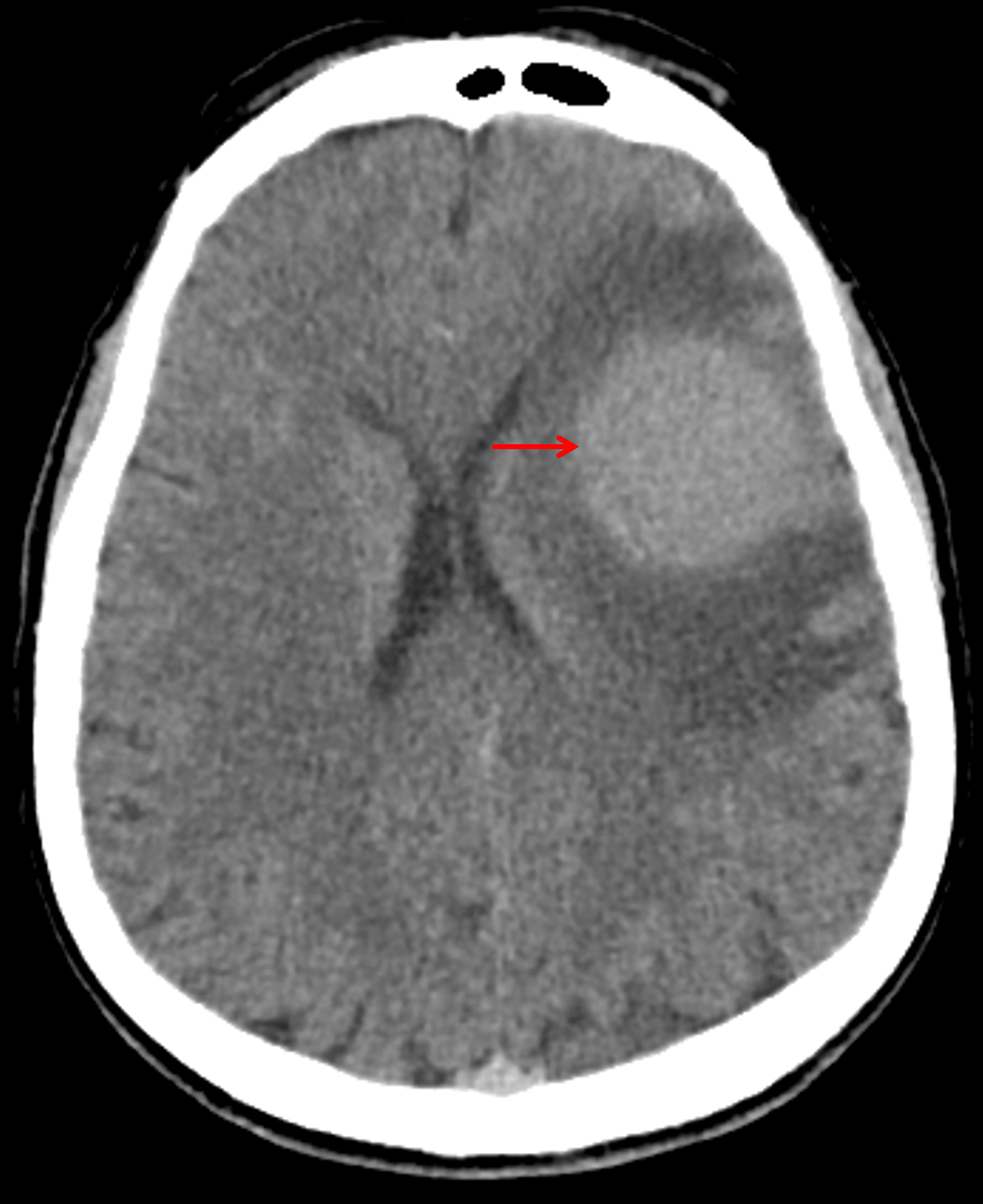
Hyperattenuating mass on CT (red arrow) with surrounding vasogenic edema.
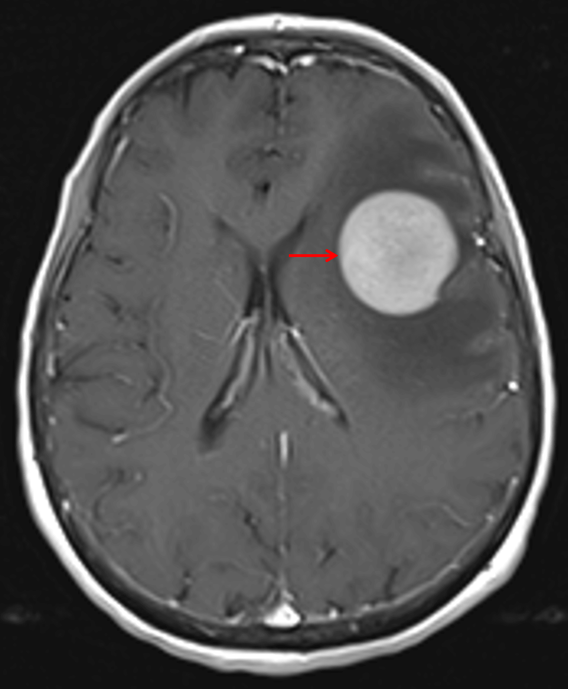
Avid, diffuse corresponding enhancement (red arrow).
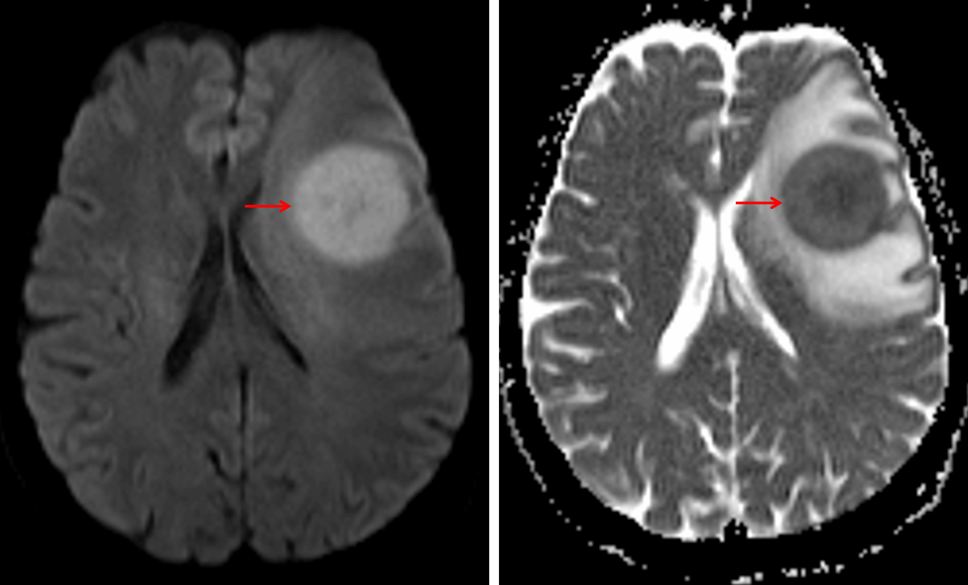
Corresponding diffuse restricted diffusion (red arrows).
Diagnosis
Primary CNS lymphoma (PCNSL)
Key Imaging Features
Become a PRO member to unlock the key imaging features
Differential Diagnosis
Become a PRO member to unlock the differential diagnosis
Discussion
Pearls
Become a PRO member to unlock the pearls