Demographics:
6 years old, Female
Indication:
Headache
Findings
- Heterogeneously enhancing mass centered in the fourth ventricle measuring 5 x 4 x 3 cm with associated diffuse restricted diffusion
- Multiple internal vessels are noted on SWI and postcontrast sequences
- Resultant effacement of the fourth ventricle and obstructive hydrocephalus with subependymal edema involving the lateral and third ventricles
- Inferior displacement of the cerebellar tonsils into the foramen magnum with crowding of the upper cervical spinal cord
Annotated Images & Illustrations
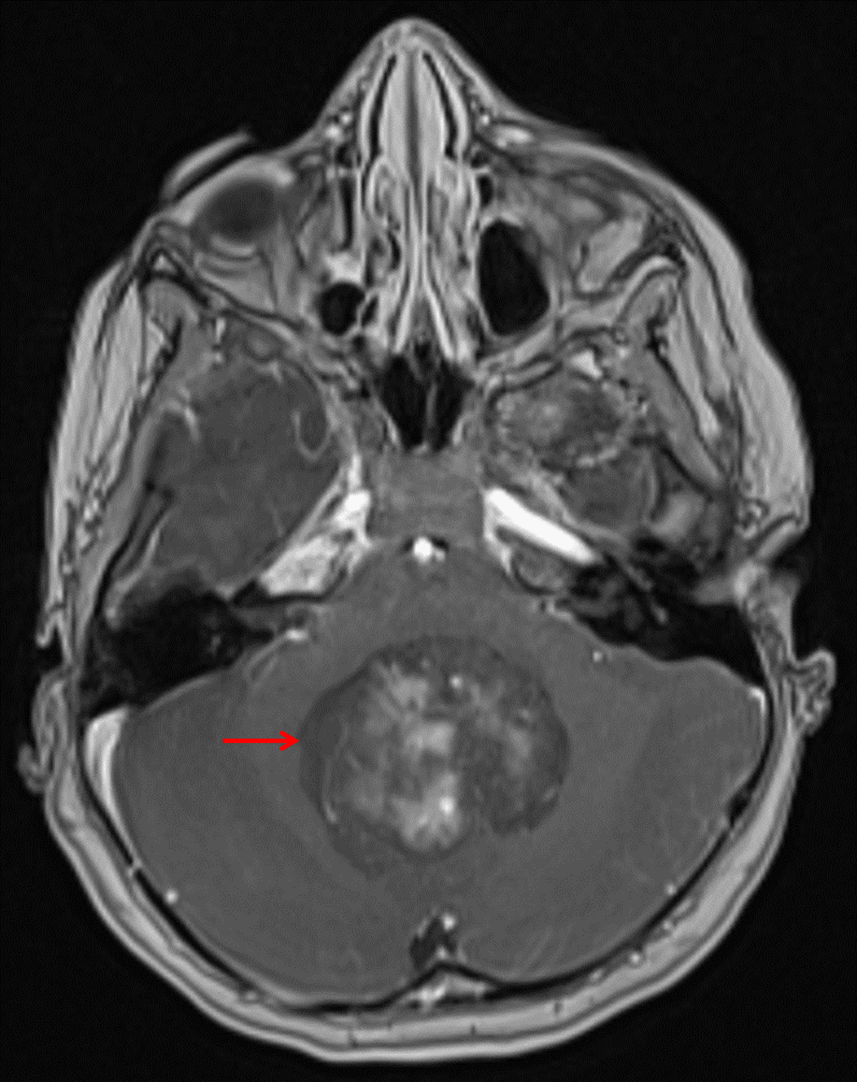
Heterogeneously enhancing mass centered in the vermis and fourth ventricle (red arrow).
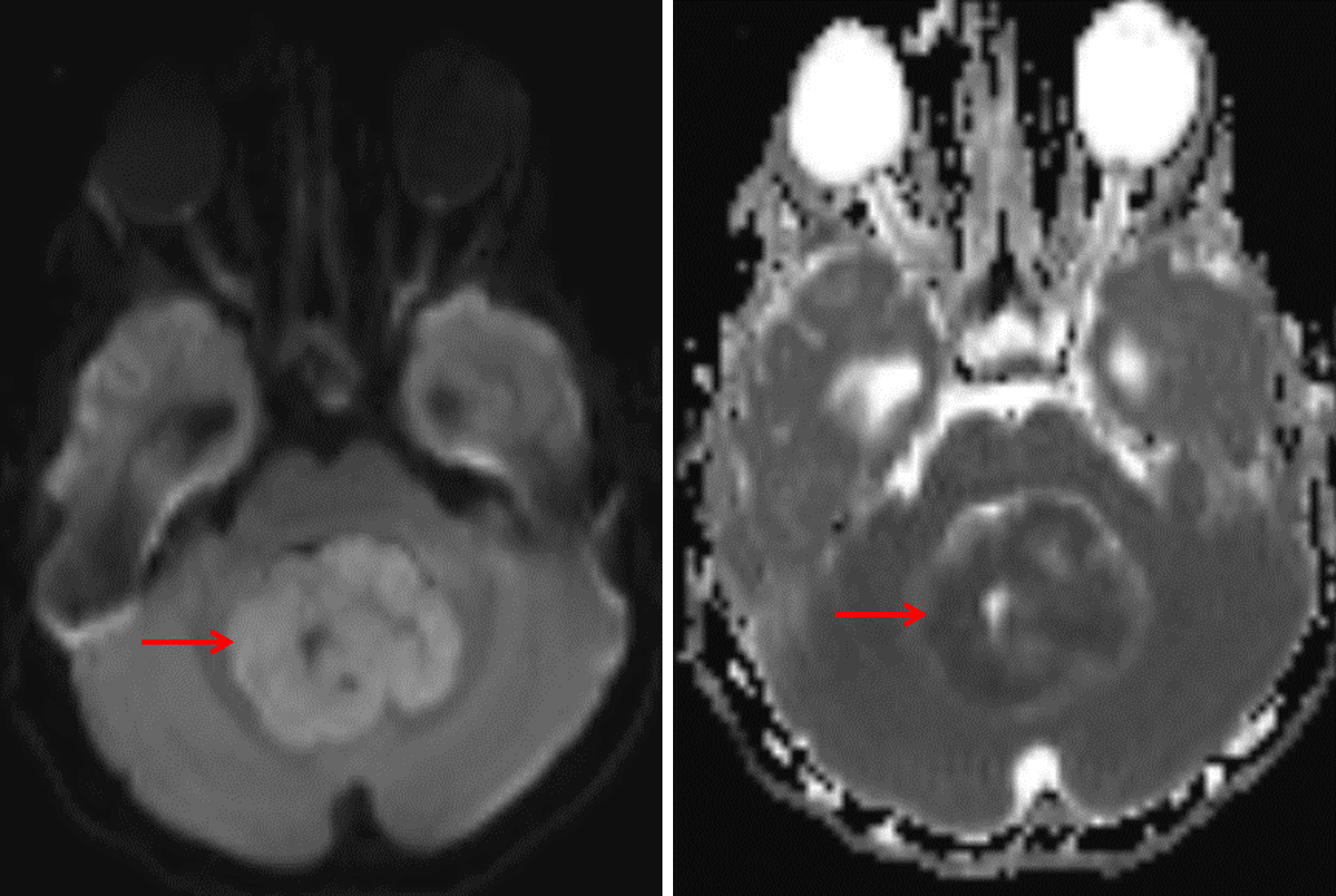
The tumor demonstrates diffuse corresponding restricted diffusion (red arrows), meaning hyperintense signal on the diffusion sequence (left) and hypointense signal on the ADC map (right).
Diagnosis
Medulloblastoma
Key Imaging Features
Become a PRO member to unlock the key imaging features
Differential Diagnosis
Become a PRO member to unlock the differential diagnosis
Discussion
Pearls
Become a PRO member to unlock the pearls