Demographics:
36 years old, Female
Indication:
Syncope, possible seizure
Findings
CT
- Ill-defined mixed hypoattenuating and hyperattenuating lesion in the inferior left temporal lobe
MRI
- Mixed cystic and solid mass in the inferior left temporal lobe measuring 1.6 x 1.5 x 1.3 cm with mild surrounding vasogenic edema
- The solid components diffusely enhance
- No corresponding restricted diffusion
- No substantial mass effect
Annotated Images & Illustrations
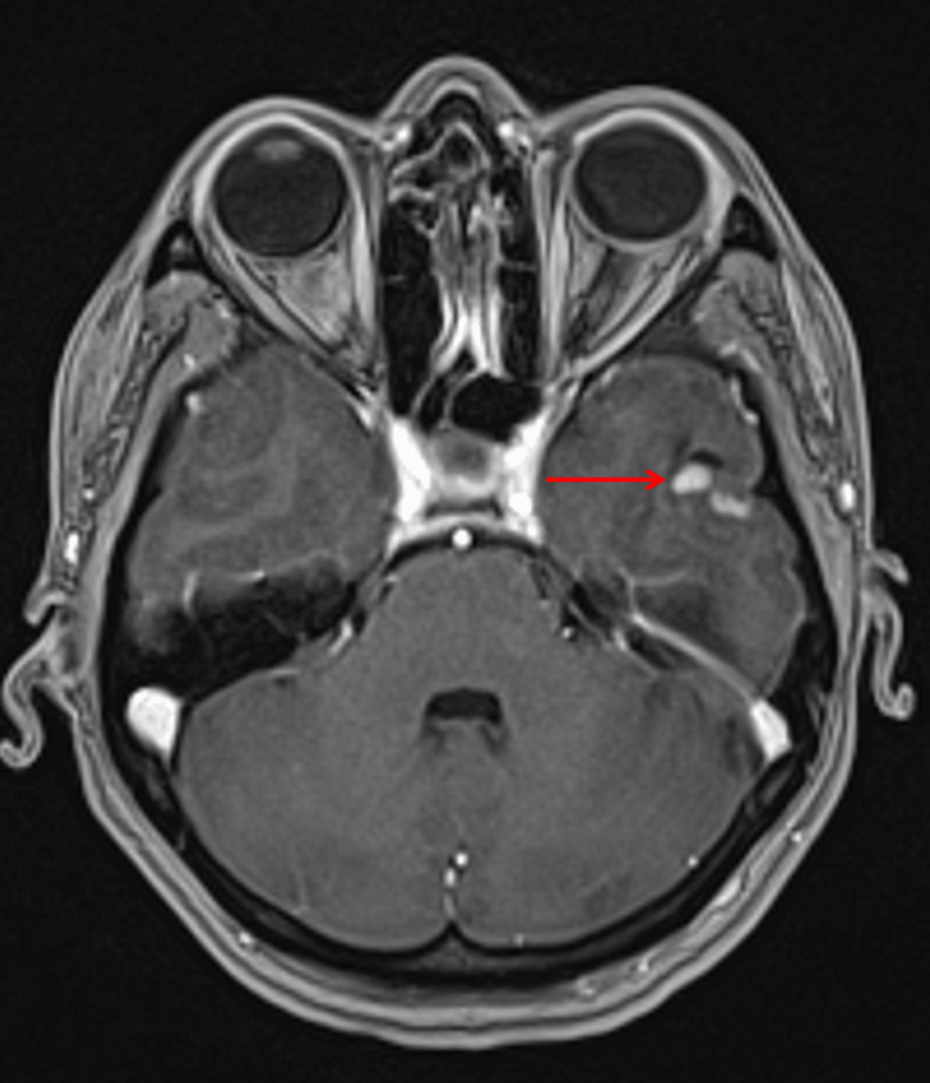
Small, mixed cystic and solid lesion in the inferior left temporal lobe with avid enhancement of the solid components (red arrow).
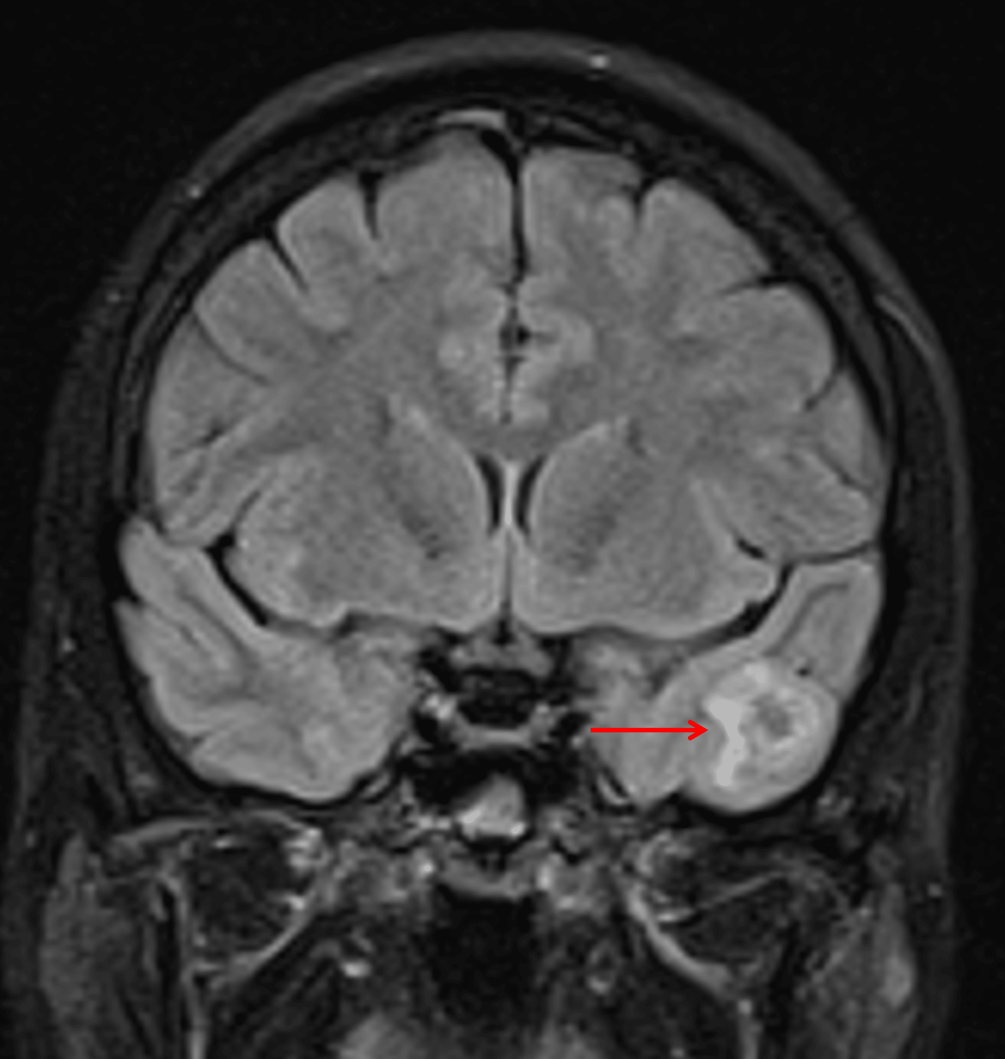
Minimal surrounding edema and mass effect (red arrow), which is typical for gangliogliomas.
Diagnosis
Ganglioglioma
Key Imaging Features
Become a PRO member to unlock the key imaging features
Differential Diagnosis
Become a PRO member to unlock the differential diagnosis
Discussion
Pearls
Become a PRO member to unlock the pearls