Findings
CT
- Hyperattenuating posterior fossa mass centered in the fourth ventricle
- Obstructive hydrocephalus with marked enlargement of the lateral and third ventricles and periventricular edema
- Generalized sulcal and cisternal effacement
MRI
- Large, heterogeneously enhancing mass arising at the floor of the fourth ventricle and extending through the right greater than left foramina of Luschka and foramen of Magendie into the prepontine and premedullary cisterns and upper cervical spinal canal
- Extensive corresponding mass effect on the cerebellum, pons, medulla, and upper cervical spinal cord
- T2 signal intensity is similar to that of gray matter
- No corresponding restricted diffusion
- A few foci of internal susceptibility artifact likely represent calcification in correlation with the CT
- Encasement of the basilar and intradural vertebral arteries
Annotated Images & Illustrations
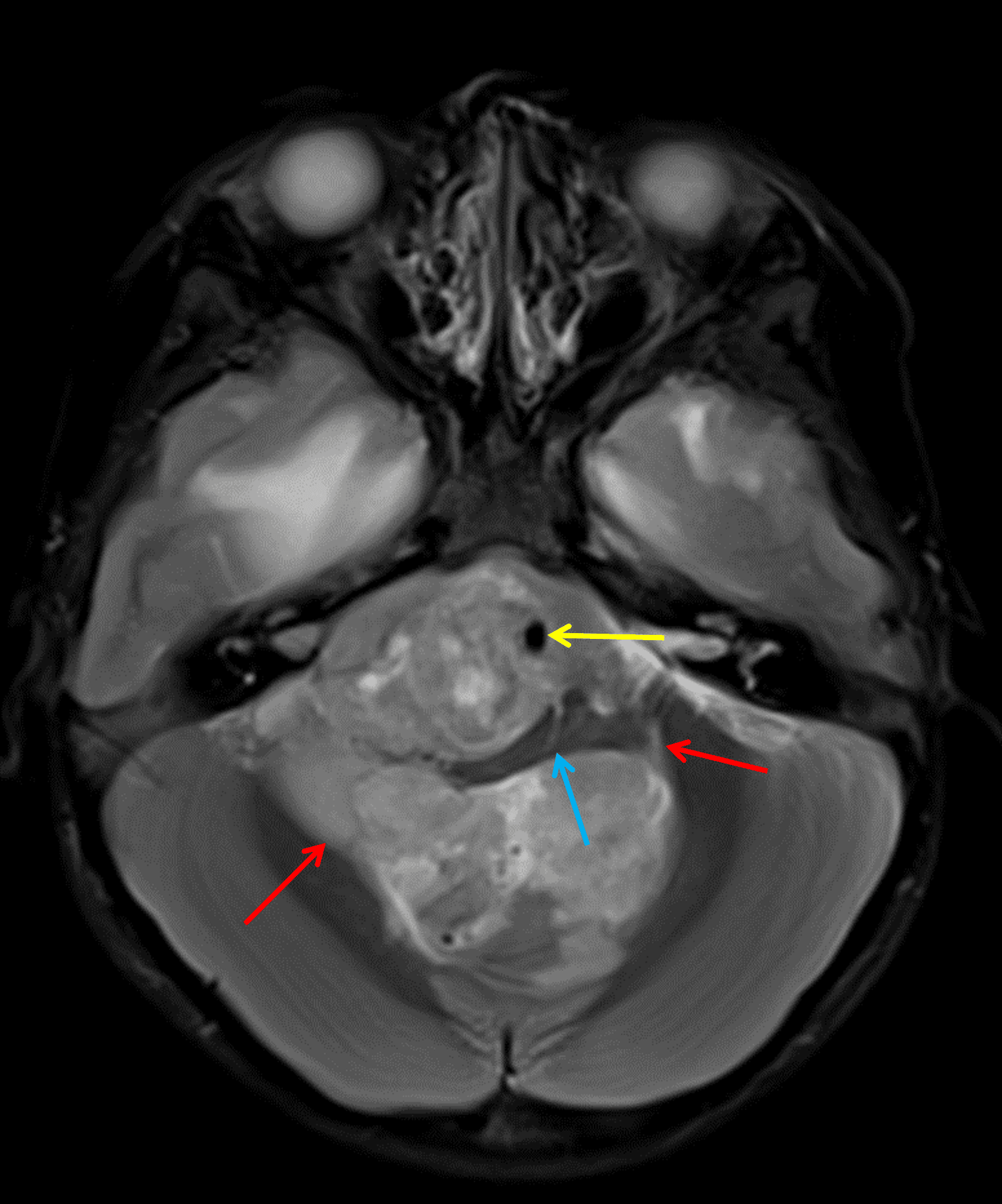
Posterior fossa ependymoma extending through the foramina of Luschka (red arrows), encasing the basilar artery (yellow arrow), and severely deforming the medulla (blue arrow).
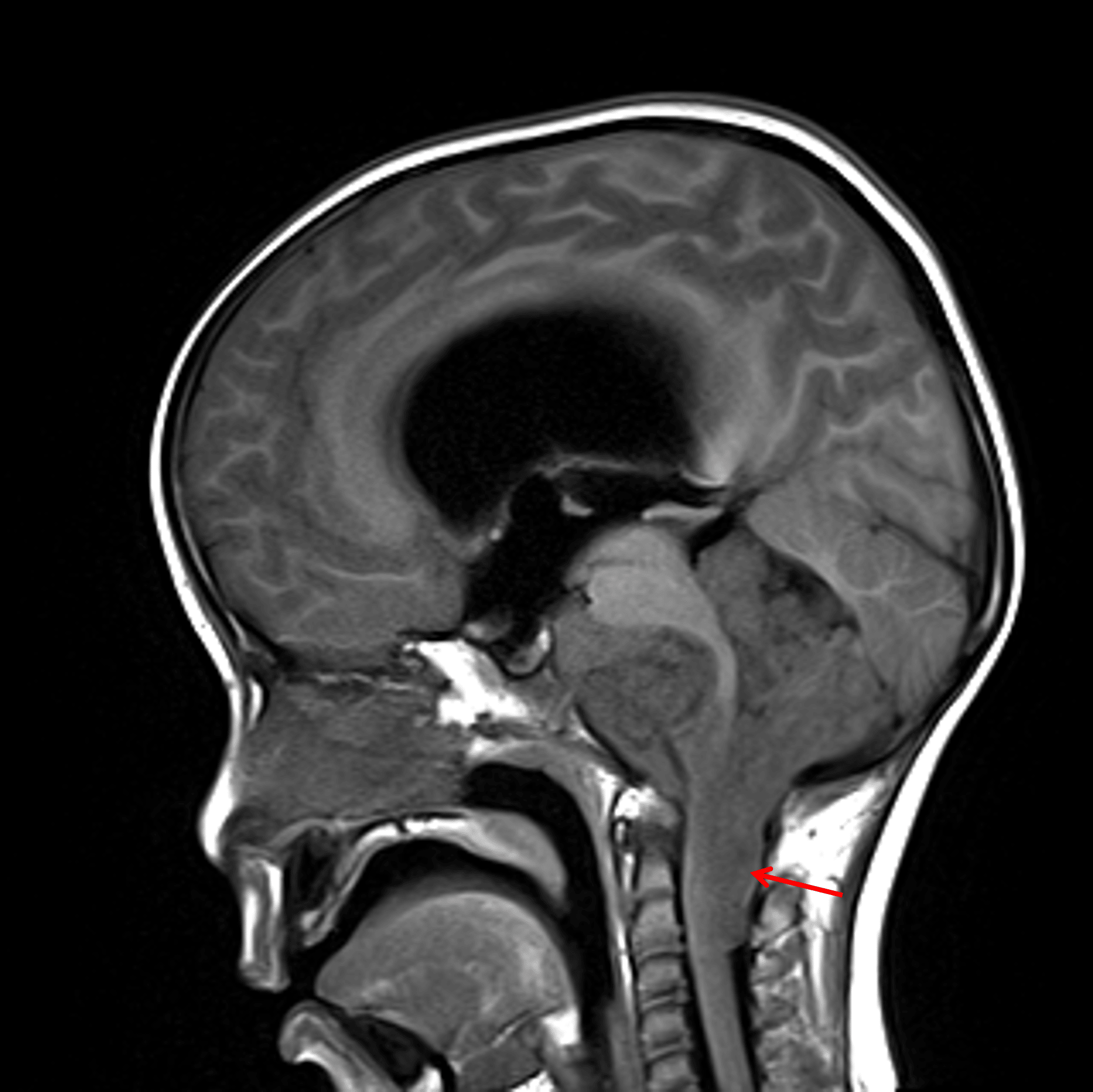
Sagittal T1 precontrast image shows the tumor encasing multiple posterior fossa structures and extending inferiorly within the dorsal aspect of the upper cervical spinal canal (red arrow).
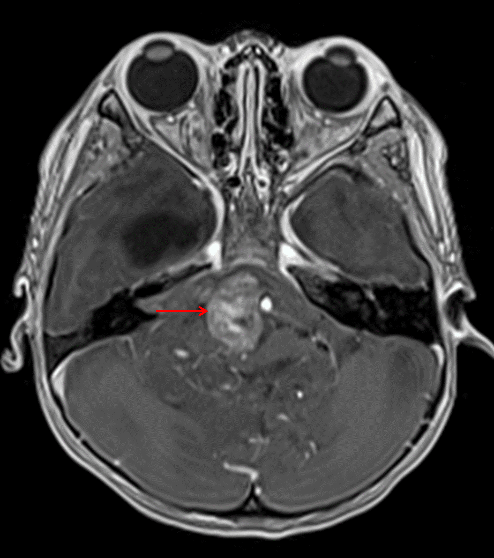
Heterogeneous areas of internal enhancement, including in the prepontine tumor component (red arrow).
Diagnosis
Ependymoma
Key Imaging Features
Become a PRO member to unlock the key imaging features
Differential Diagnosis
Become a PRO member to unlock the differential diagnosis
Discussion
Pearls
Become a PRO member to unlock the pearls