マウスホイール、矢印キー、または左クリック(スクロールツール使用時)でスクロールしてください。
1本の指でドラッグ(スクロールツール使用時)してスクロールしてください。
DICOMヘルプSource: Local (us-east1-c)
患者背景:
34 歳, 男性
適応:
肩の外傷
所見
- 後方肩関節脱臼
- 上腕骨頭前方の急性逆Hill-Sachs陥没骨折
- 後方関節窩の急性軽度転位逆Bankart骨折
診断
逆Hill-Sachs骨折およびBankart骨折を伴う後方肩関節脱臼
サンプルレポート
急性逆Hill-Sachs陥没骨折および急性軽度転位逆Bankart病変を伴う後方肩関節脱臼。
肩鎖関節のアライメントは保たれている。
考察
- 肩関節脱臼には前方、後方、下方の3つの主要な種類があり、前方が圧倒的に最も一般的である。各種脱臼のX線写真上の手がかりを以下に示す:
- 前方:上腕骨頭が関節窩の下方かつ内側に位置
- 後方:正面像では上腕骨頭が関節窩に対して正常に見えることがある。肩は典型的に内旋位に固定される(いわゆる"電球"サイン)
- 下方:上腕骨頭が関節窩の下方に位置し、腕がしばしば外転位に固定(頭上に挙上、"luxatio erecta")
- 関連骨折:
- Hill-Sachs
- 上腕骨頭が関節窩に衝突する上腕骨頭後外側に沿って発生
- 内旋像で最もよく見える
- 後方脱臼による逆Hill-Sachs -> 上腕骨頭前内側の陥没骨折、X線写真上"トラフ(溝)"サインとして現れる
- Bankart
- 関節窩前縁、X線写真では検出困難なことが多い
- 後方肩関節脱臼では逆Bankart(後縁)
- Hill-Sachs
注釈付き画像とイラスト
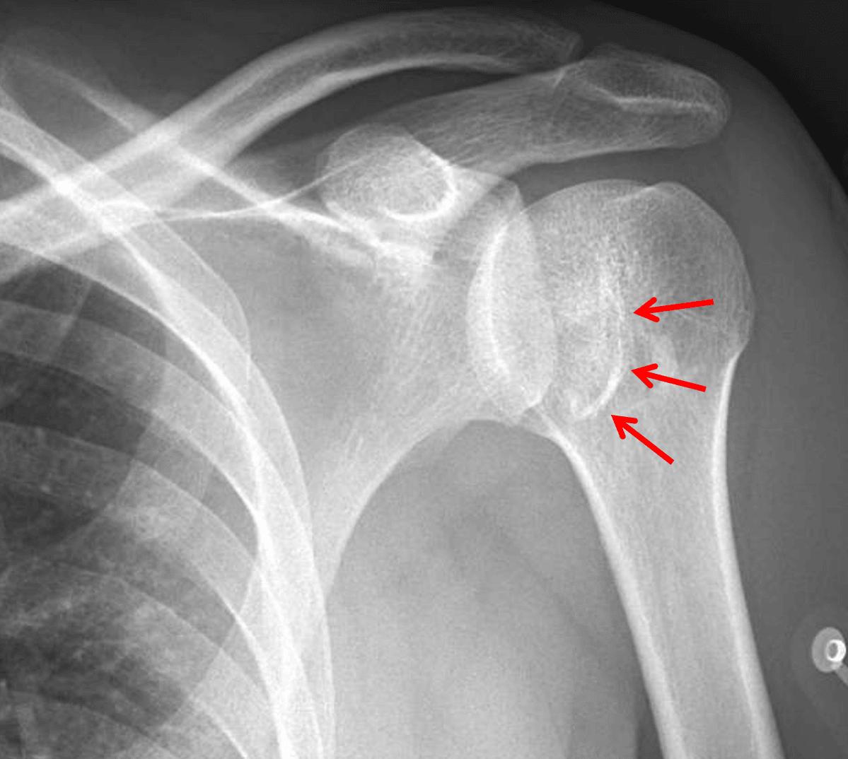
Red arrows: reverse Hill-Sachs fracture of the inferomedial humeral head resulting in a trough sign on frontal views.
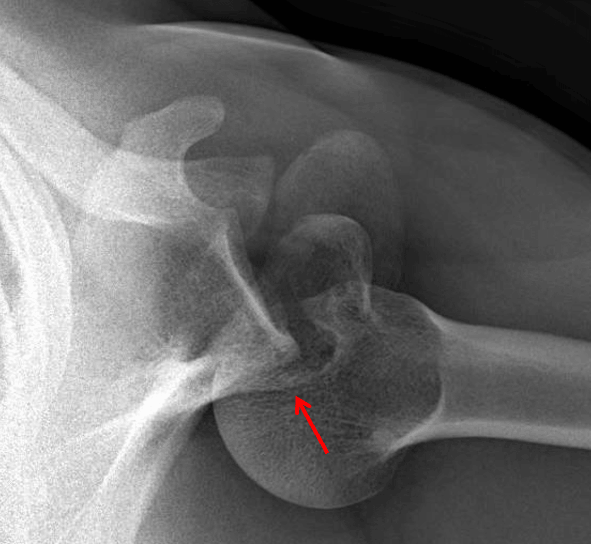
Red arrow: posterior shoulder dislocation with minimally displaced fracture of the posterior glenoid, best shown on this axillary view.