マウスホイール、矢印キー、または左クリック(スクロールツール使用時)でスクロールしてください。
ui.case.use_touch_gestures
DICOMヘルプSource: Local (us-east1-c)
症例 #1
所見
診断
右上葉無気肺
サンプルレポート
右上葉無気肺。粘液栓または中枢性閉塞性腫瘤が懸念される。精査のため胸部CTを推奨する。
左下葉の軽度斑状不透過化は非特異的であり、無気肺、誤嚥、または肺炎に関連する可能性がある。
考察
- 右上葉無気肺は正面レントゲンで小葉間裂の上方偏位を伴う容量減少と不透過化により同定されることが多い
- Golden S signは小葉間裂の内側部分が肺門腫瘤に引っかかり、中央部および外側部が上方に偏位することで生じるため、この外観を認めたら中枢性閉塞性腫瘤を考えること
注釈付き画像とイラスト
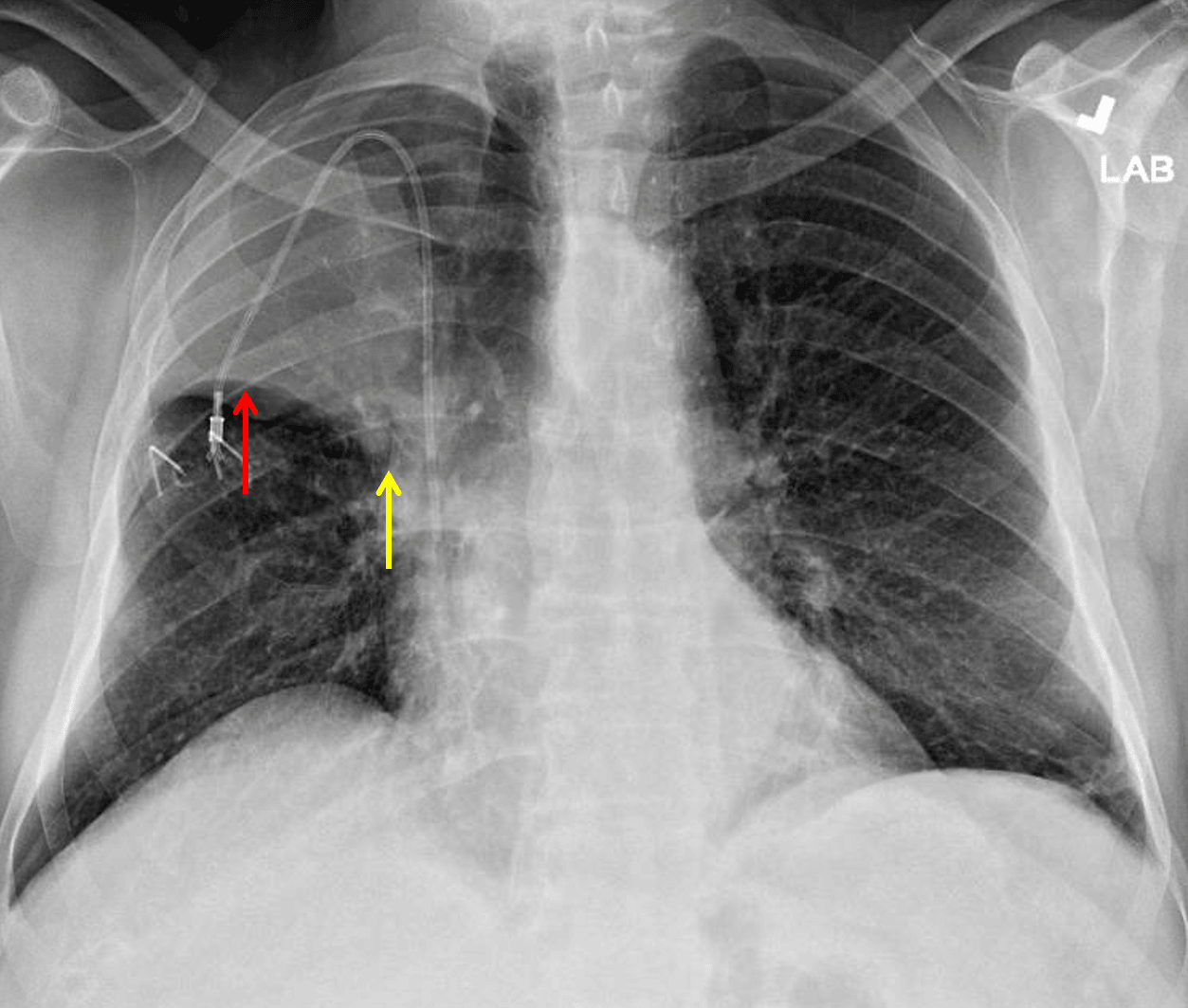
Right upper lobe collapse. The minor fissure is elevated laterally (red arrow) but not as elevated centrally (yellow arrow) raising concern for a perihilar mass.
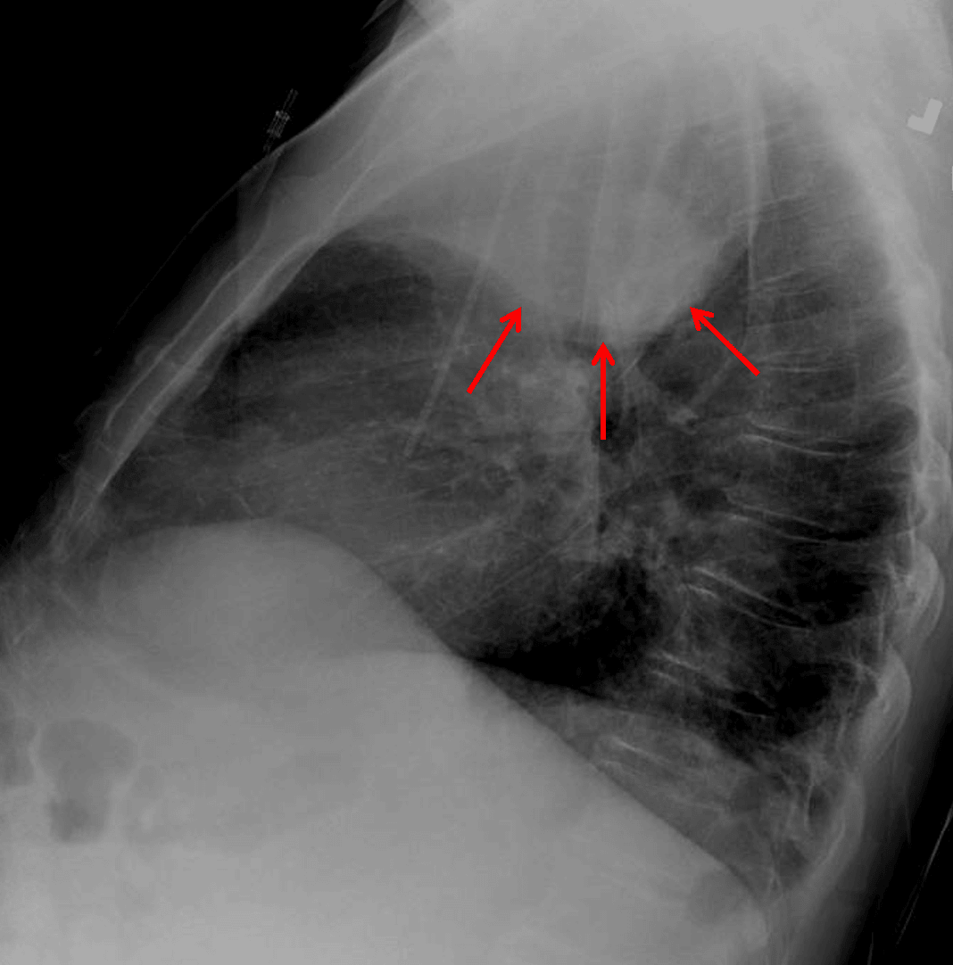
Right upper lobe collapse. Lateral view shows a paradoxical bulging appearance along the inferior margin of the collapsed lobe concerning for mass (red arrows).
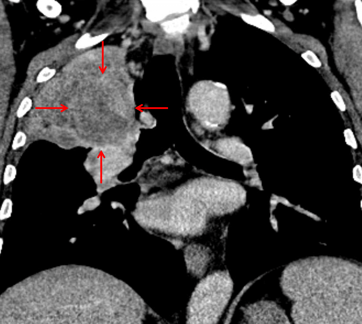
Subsequent CT confirmed the presence of a suprahilar mass with bronchial invasion (red arrows), though the mass is difficult to distinguish from the adjacent atelectatic lung.
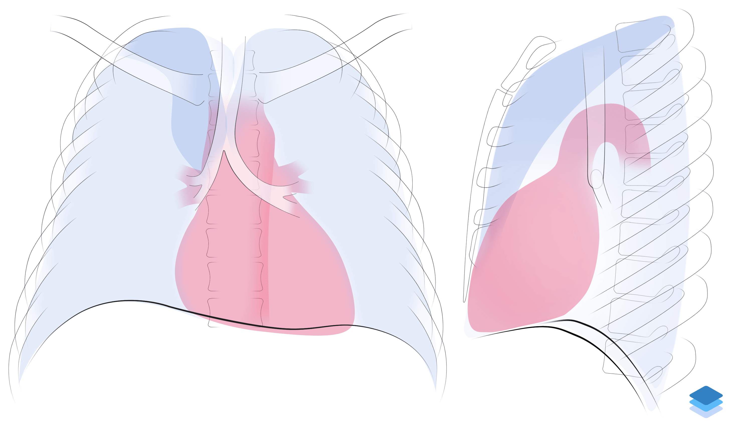
Right upper lobe collapse. Illustration by Valerie George, MD