Use mouse wheel, arrow keys or left click (with scroll tool selected) to scroll
ui.case.use_touch_gestures
DICOM HelpSource: Local (us-east1-c)
Findings
-
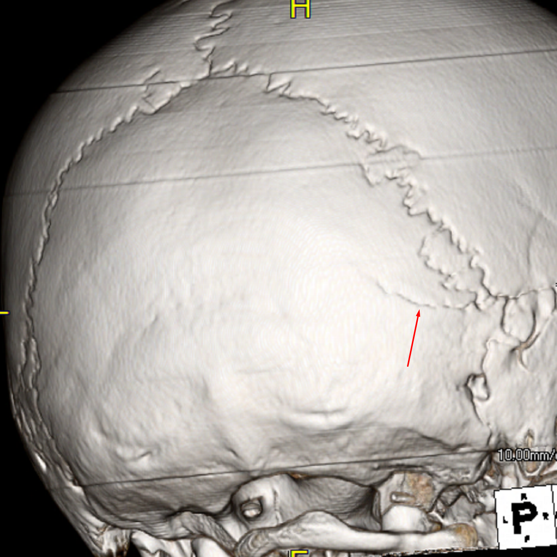
Non-depressed zigzag lucency involving the right occipital bone, which is located medial to the lambdoid suture and superior to the occipitomastoid suture. No diastasis. Given the appearance and location, this finding is consistent with a persistenet mendosal suture rather than an acute fracture.
- No overlying soft tissue contusion and no intracranial hemorrhage.
Diagnosis
Mendosal Suture
Discussion
-
The mendosal suture (aka the accessory occipital suture) usually closes well before birth, but it can persist into childhood and very rarely adulthood.
-
The suture is located in the occipital bone. On axial images it is seen medial to the lambdoid suture and on coronal images it is located superior to the occipitomastoid suture.
-
This suture is easy to misdiagnose as a fracture and is often unilateral (as in this case). Knowing the normal location of the mendosal suture is key. Use your 3D reformatted imaging to help you.
-
There also are some general principles that can help you differentiate suture from fracture. Sutures tend to join other sutures (not cross them) and are more likely to have a "zigzag" appearance. Fractures may cause diastasis (widening) of sutures, cross sutures, cause depression of fracture fragments, and generally have non-sclerotic margins and a more linear (versus "zigzag") in appearance.
-
Look for any overlying soft tissue contusion or subjacent extra-axial hemorrhage to increase your suspicion for fracture.
-
Of note, this patient also has a partially fused metopic suture best seen near the bregma.
-
Annotated Images & Illustrations
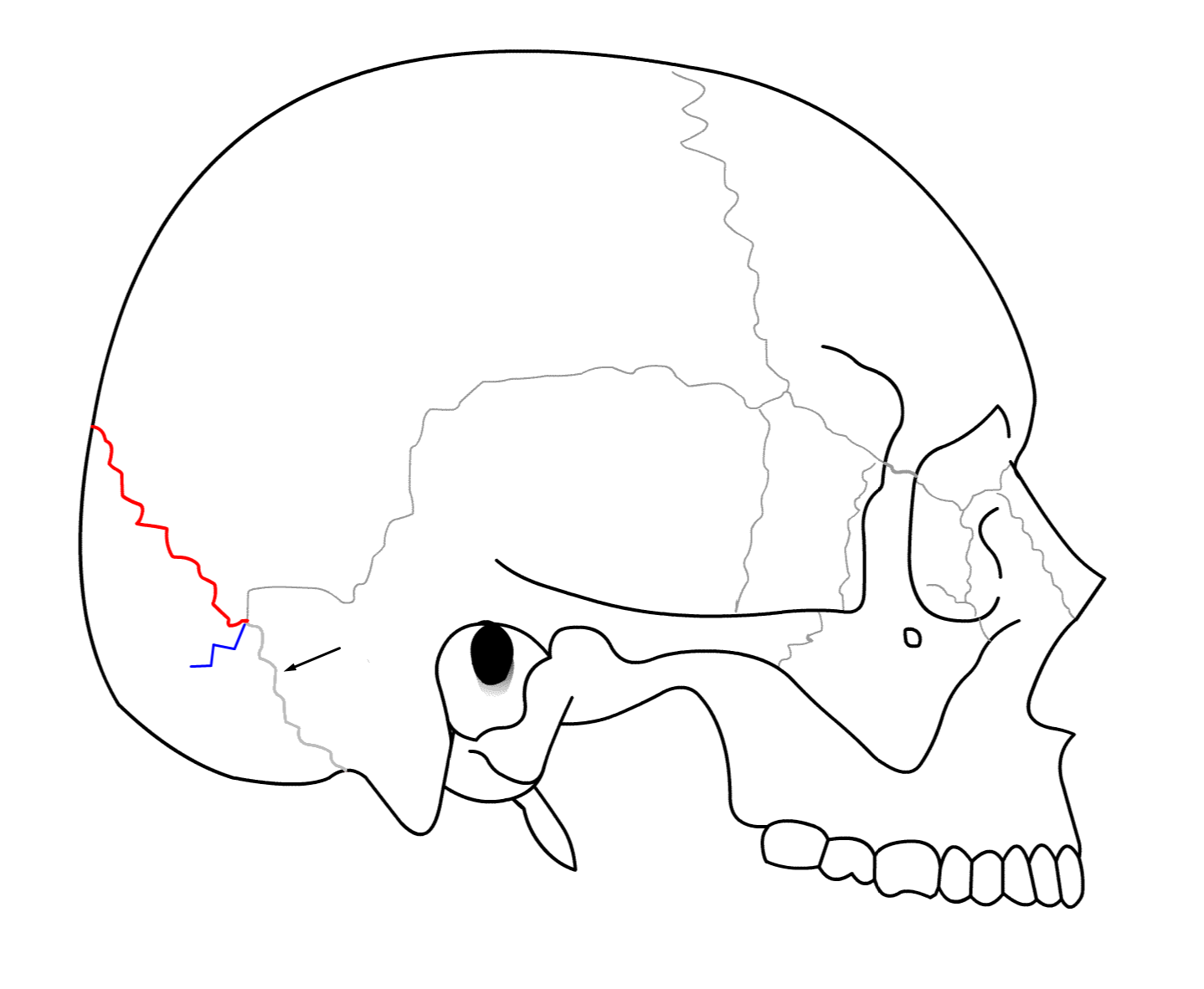
mendosal-illustration
mendosal
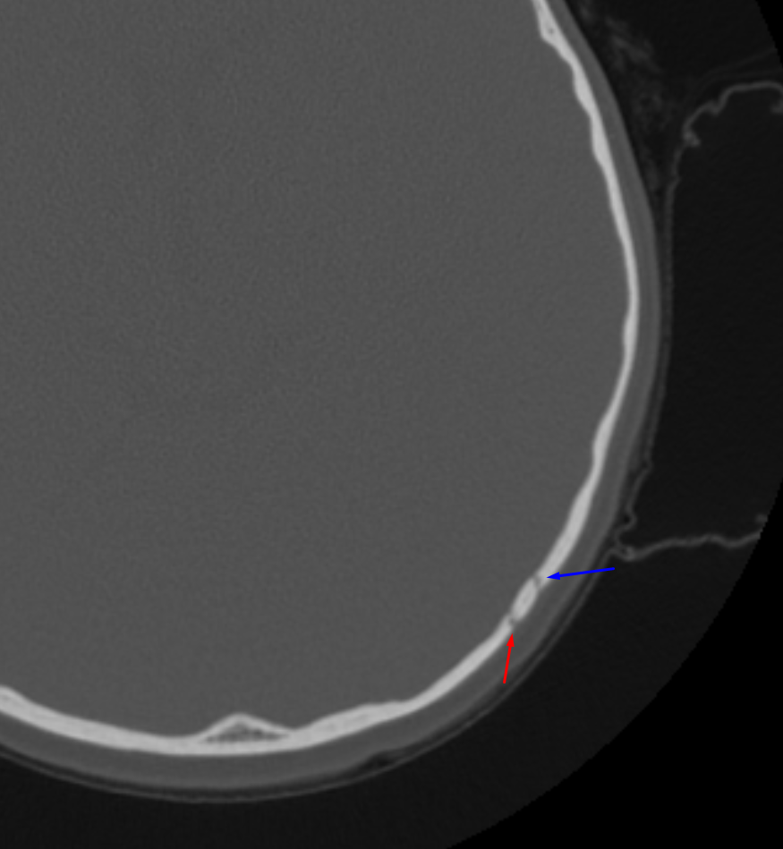
mendosal-axial
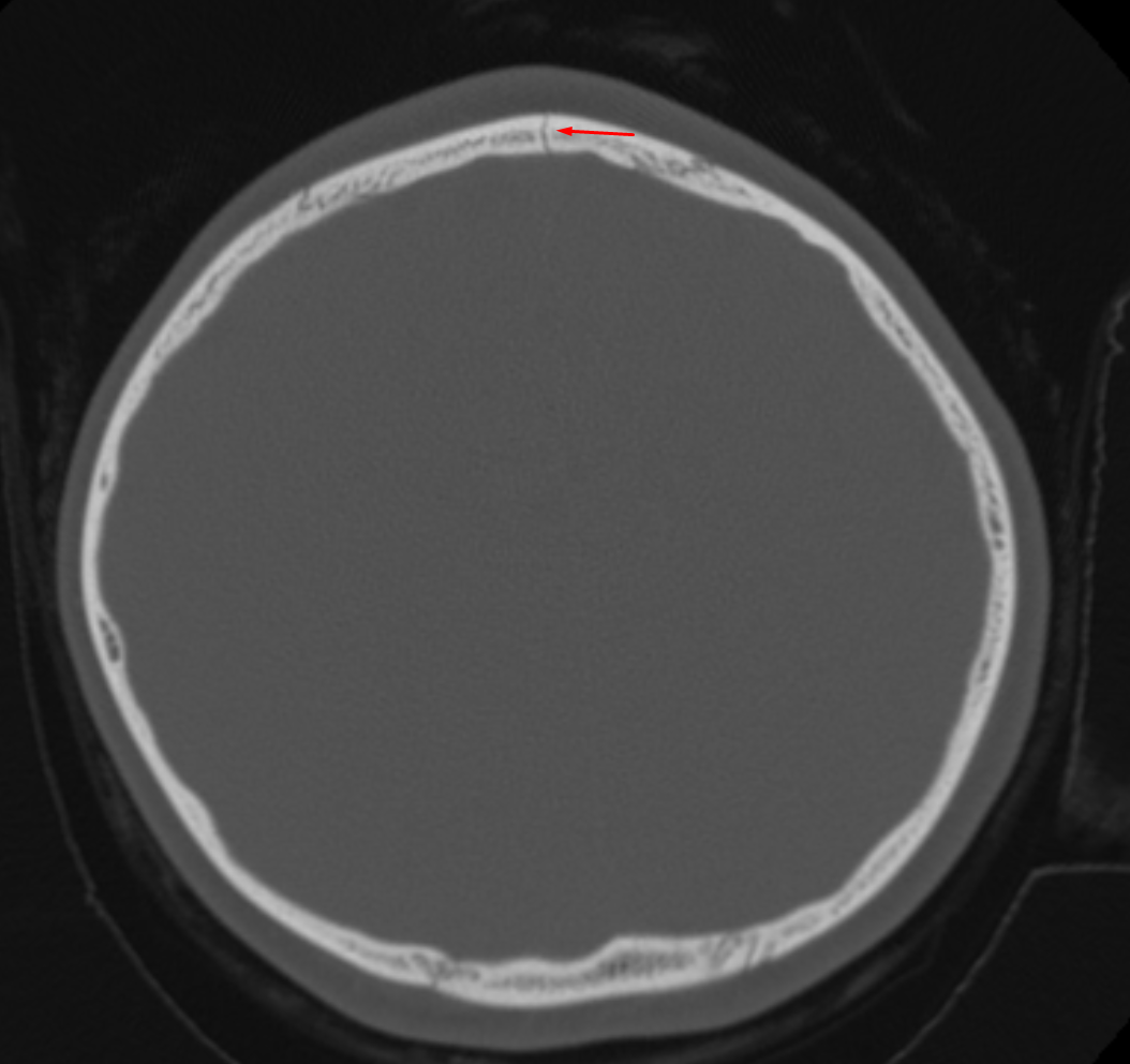
metopic-2