Use mouse wheel, arrow keys or left click (with scroll tool selected) to scroll
ui.case.use_touch_gestures
DICOM HelpSource: Local (us-east1-c)
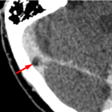
Findings
- Small rounded filling defect within the right transverse sinus, compatible with an arachnoid granulation.
Diagnosis
Arachnoid Granulation Mimicking Thrombus
Discussion
-
Arachnoid granulations are common, with a prevalence of approximately 60%.
-
They are protrusions of arachnoid through the dura mater, which allow CSF to extend from the subarachnoid space into the venous sinus or into the calvarium.
-
They most commonly occur in the superior sagittal and transverse sinuses.
-
On non-contrast head CT they are CSF density and on post-contrast imaging they appear as round, focal filling defect in the dural sinus. Usually they are small (<10mm), but occassionally can be "giant" (>10mm). If they involve the calvarium, they appear as a circumscribed lucent lesion.
-
Usually these granulations are easy to differentiate from thrombosis given their round, well-circumscribed appearance and typical location.
-
Annotated Images & Illustrations
arachnoid granulation