Use mouse wheel, arrow keys or left click (with scroll tool selected) to scroll
ui.case.use_touch_gestures
DICOM HelpSource: Local (us-east1-c)
Findings
- Displaced left occipital condyle fracture with widening and posterior offset of the right atlanto-occipital articulation. Displaced left occipital condyle fracture fragments narrow the anterior and left aspects of the foramen magnum
- Right 1st and 2nd rib fractures with right greater than left apical pulmonary opacification
- Partially imaged endotracheal tube
Diagnosis
Atlanto-occipital dislocation
Sample Report
Atlanto-occipital fracture/dislocation with a displaced left occipital condyle fracture and widening and posterior offset of the right atlanto-occipital articulation. Displaced left occipital condyle fracture fragments narrow the anterior and left aspects of the foramen magnum. Consider MRI without contrast to further evaluate the extent of ligamentous injury and to assess for cord trauma.
Right 1st and 2nd rib fractures with right greater than left apical pulmonary contusion and/or aspiration.
Discussion
- Watch out for craniocervical junction injuries, which can occur without fractures
- Refer to the annotated images below for a demonstration of important measurements and upper limit of normal values in adults and children
- Unfortunately, soft tissue algorithm sequences are not available for this case, but if you squint you can make out some extra-axial hemorrhage associated with the fractures (see annotated images)
Annotated Images & Illustrations
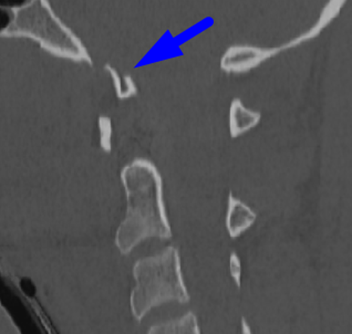
Blue arrow: displaced occipital condyle fracture.
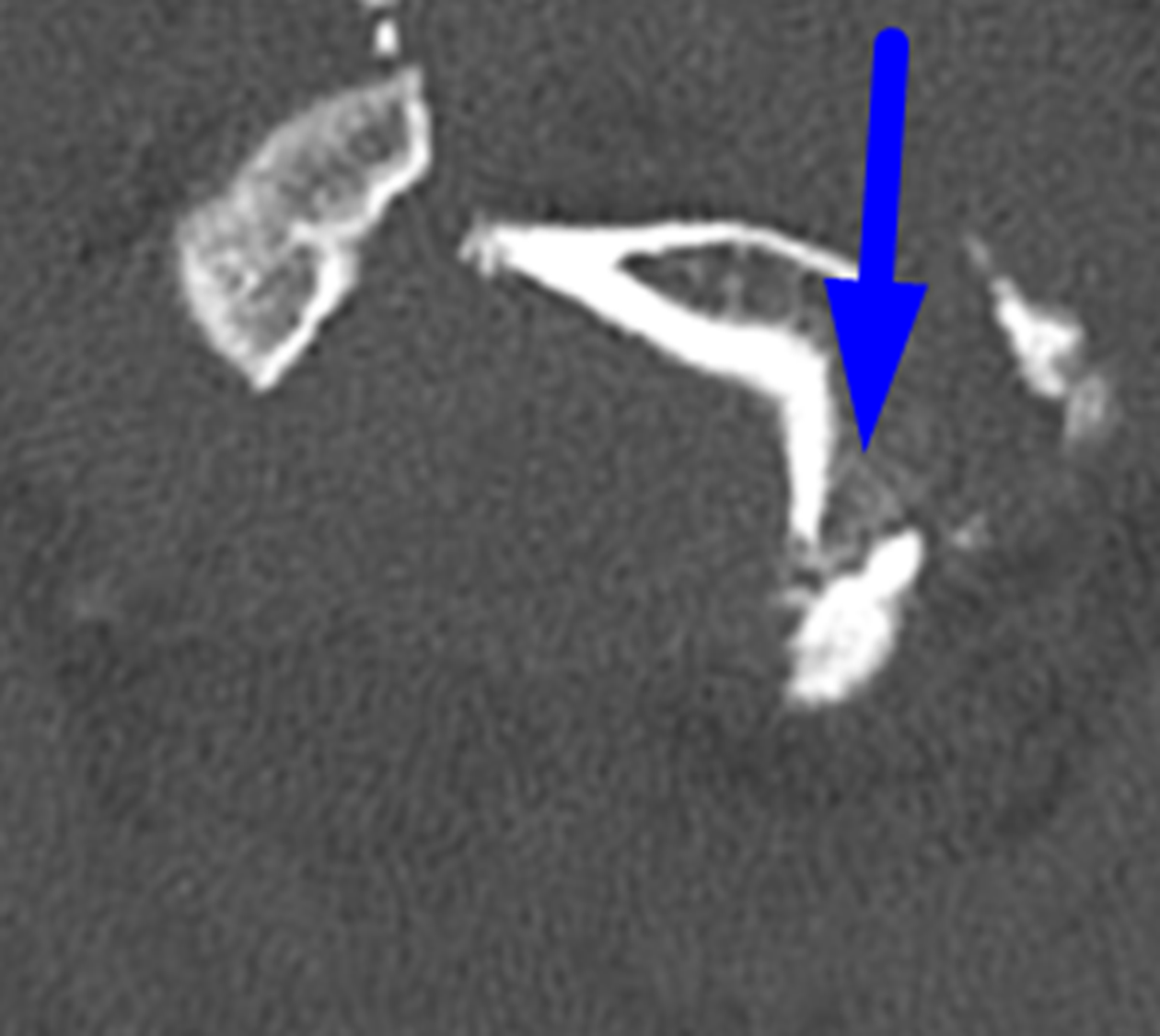
Blue arrow: displaced occipital condyle fracture.
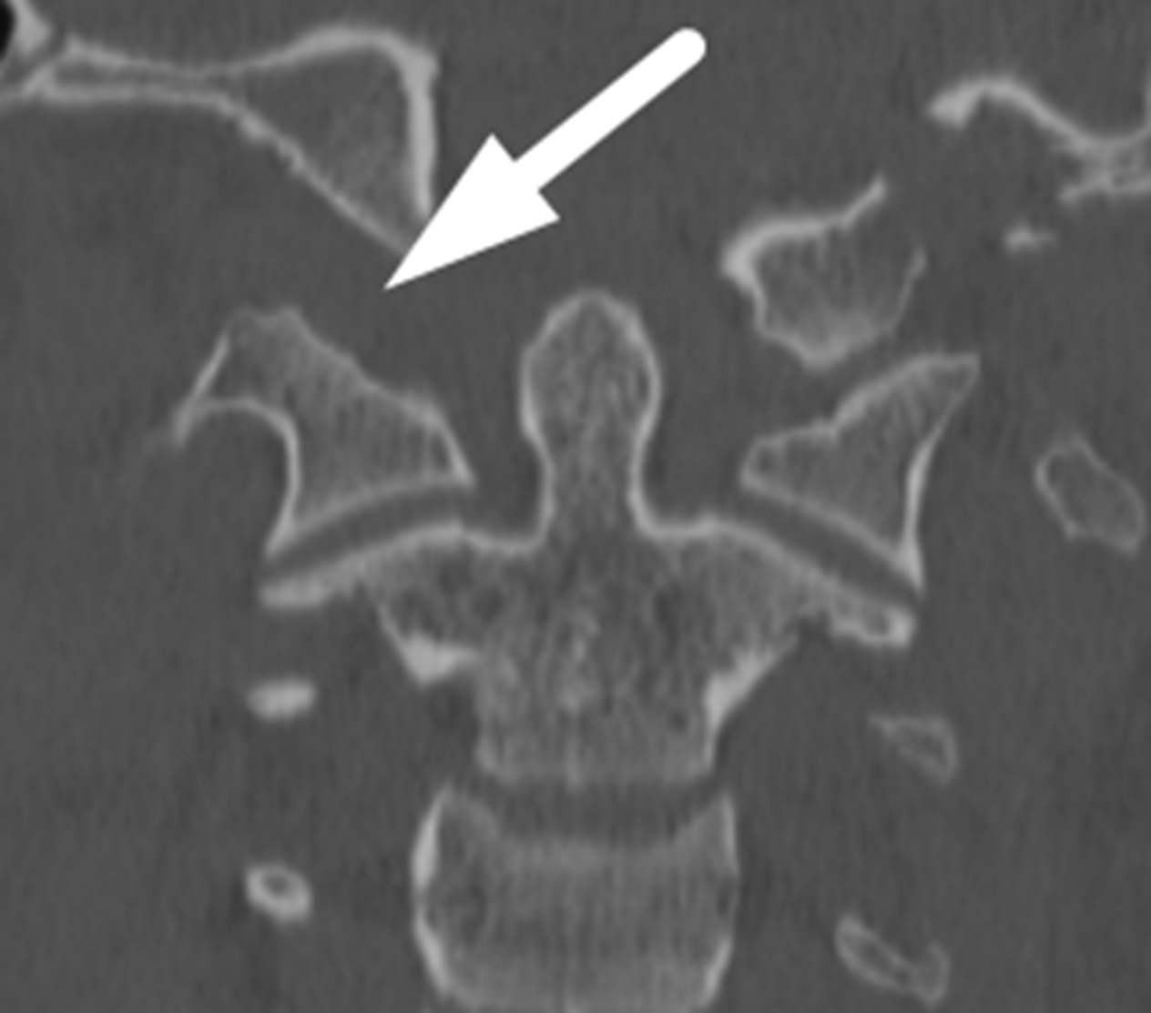
White arrow: widening of the right atlanto-occipital articulation.
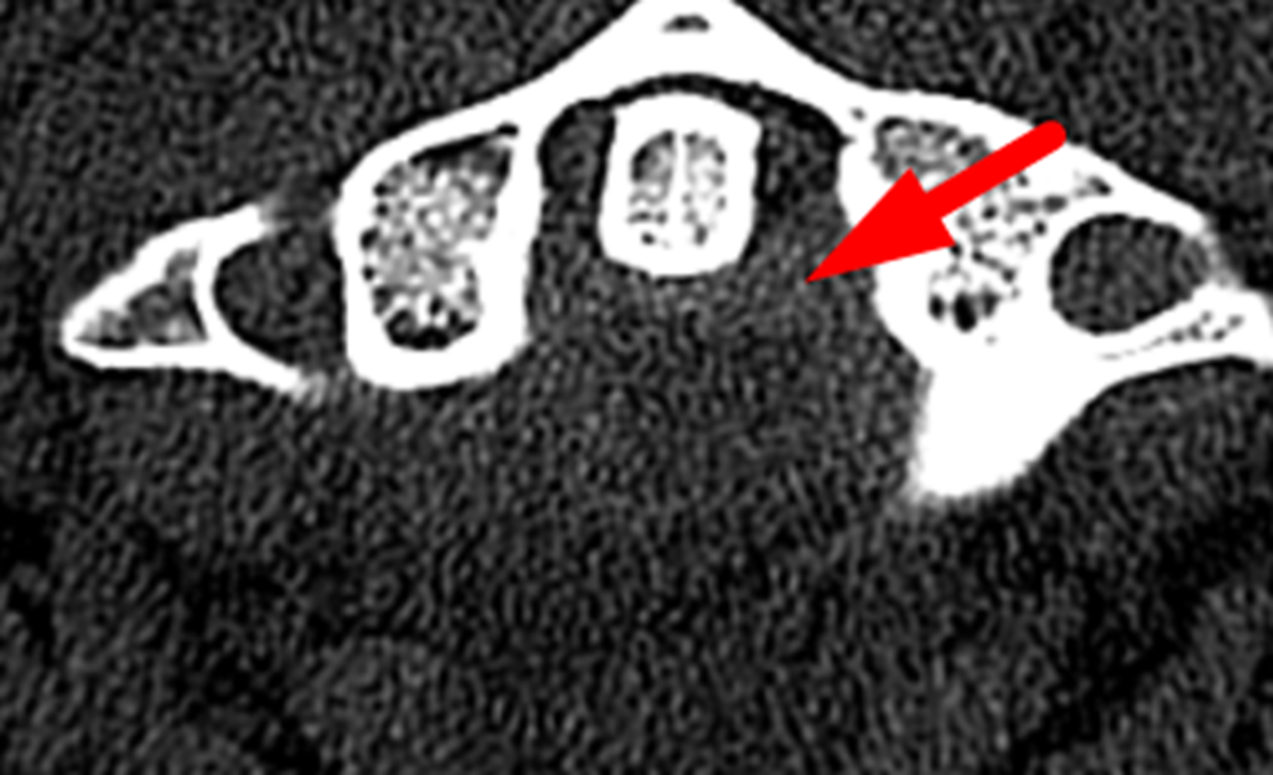
Red arrow: extra-axial hemorrhage posterior to the odontoid process.
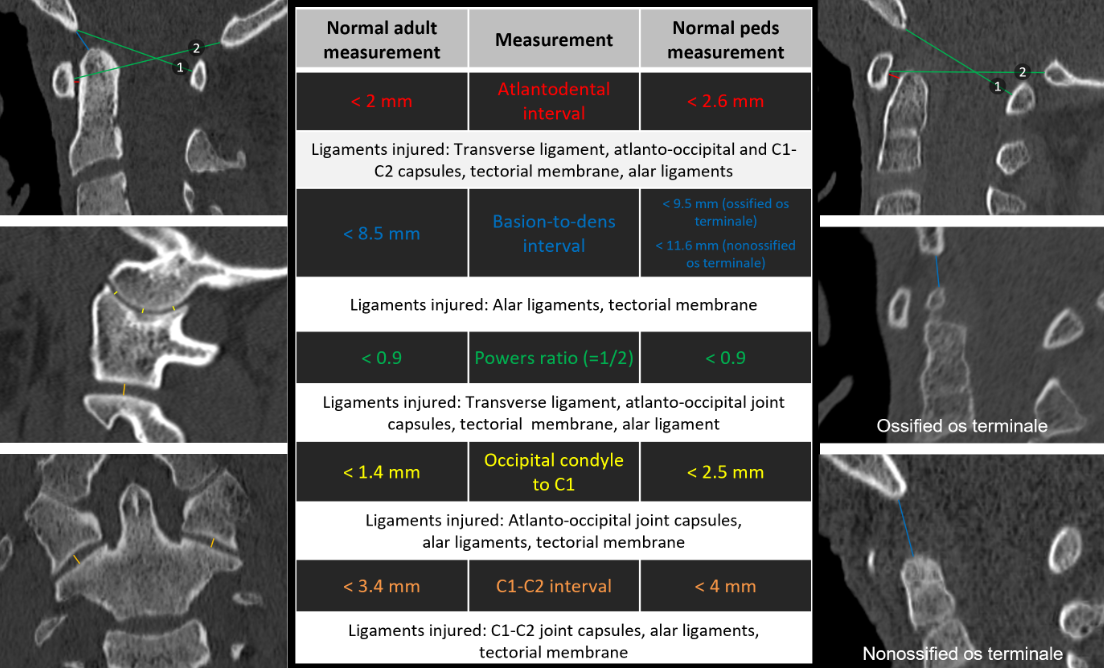
Diagram of helpful measurements to know.
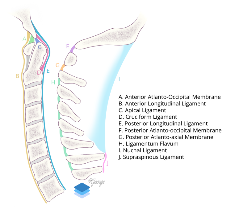
Sagittal view of the major cervical spine ligaments.
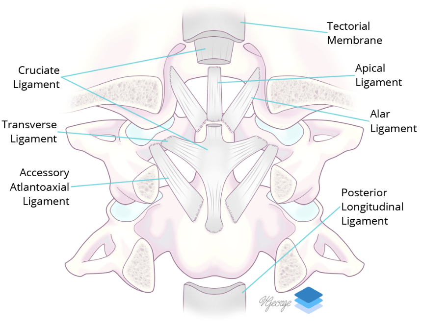
Coronal view of the major ligaments at the craniocervical junction.
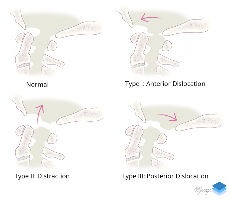
Types of craniocervical dislocation.
Related Video
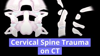
Cervical Spine Trauma
YouTube
References