Use mouse wheel, arrow keys or left click (with scroll tool selected) to scroll
ui.case.use_touch_gestures
DICOM HelpSource: Local (us-east1-c)
Findings
- Acute oblique distal fibular fracture with one-half shaft width lateral displacement and inferomedial extension above the level of the distal tibiofibular syndesmosis
- Acute medial malleolar avulsion fracture, which maintains its relationship with the talus
- Acute minimally displaced posterior malleolar fracture with mild impaction of the posterior cortex of the distal tibial metaphysis
- Approximately 50% lateral subluxation at the tibiotalar joint with medial clear space widening measuring 2.3 cm
- Subcutaneous and intraarticular gas
- Mild calcaneal enthesopathy
Diagnosis
Open Weber type C ankle fracture
Sample Report
Open acute Weber type C ankle fracture with the following components:
Acute oblique distal fibular fracture with one-half shaft width lateral displacement and inferomedial extension above the level of the distal tibiofibular syndesmosis.
Acute medial malleolar avulsion fracture, which maintains its relationship with the talus.
Acute minimally displaced posterior malleolar fracture with mild impaction of the posterior cortex of the distal tibial metaphysis.
Approximately 50% lateral subluxation at the tibiotalar joint with medial clear space widening measuring 2.3 cm, concerning for injuries to the distal tibiofibular syndesmosis and deltoid ligament.
Discussion
- While more complex ankle fracture classification systems exist, it is important to at least be familiar with the Weber classification:
- Weber type A: lateral malleolar fracture below the level of the talar dome – these are typically stable and managed conservatively
- Weber type B: lateral malleolar fracture with fracture line exiting medially at the level of the talar dome – these are stable if the medial structures are intact: look for widening of the medial clear space (indicating deltoid ligament injury) and widening of the distal tibiofibular syndesmosis
- Weber type C: lateral malleolar fracture above the level of the talar dome – usually unstable as these are associated with injury to the distal tibiofibular syndesmosis
- Just like the radius and ulna, think about the tibia and fibula as a ring structure. If you see injury in one place (either fracture or joint subluxation) inspect the rest of the ring for additional injuries. If you only see widening of the medial clear space of the ankle mortise, make sure to get tibia/fibula radiographs to look for proximal fibular (Maisonneuve) fracture
Annotated Images & Illustrations
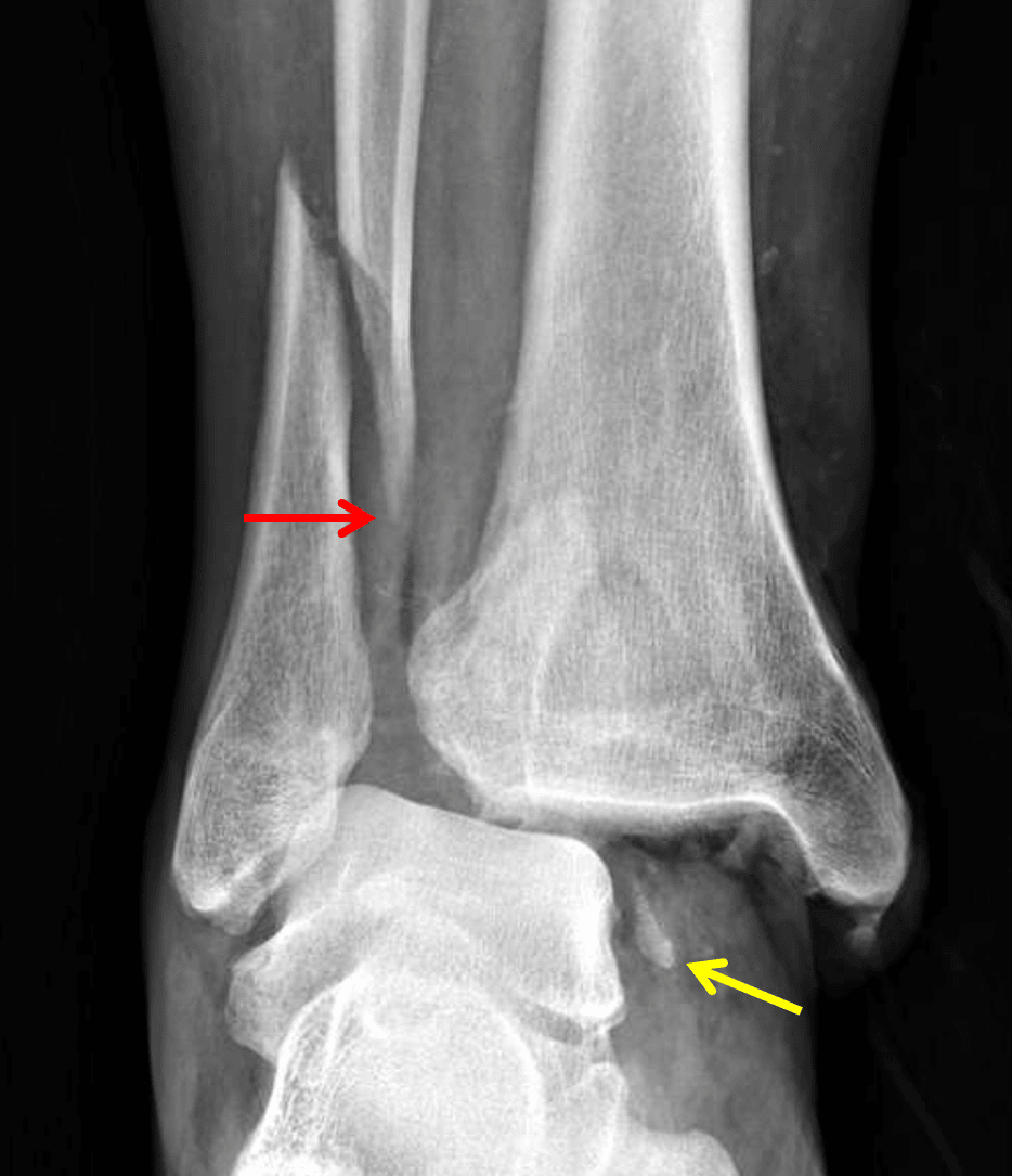
Weber type C fibular fracture (red arrow). Medial malleolar avulsion fracture remains associated with the talus (yellow arrow).
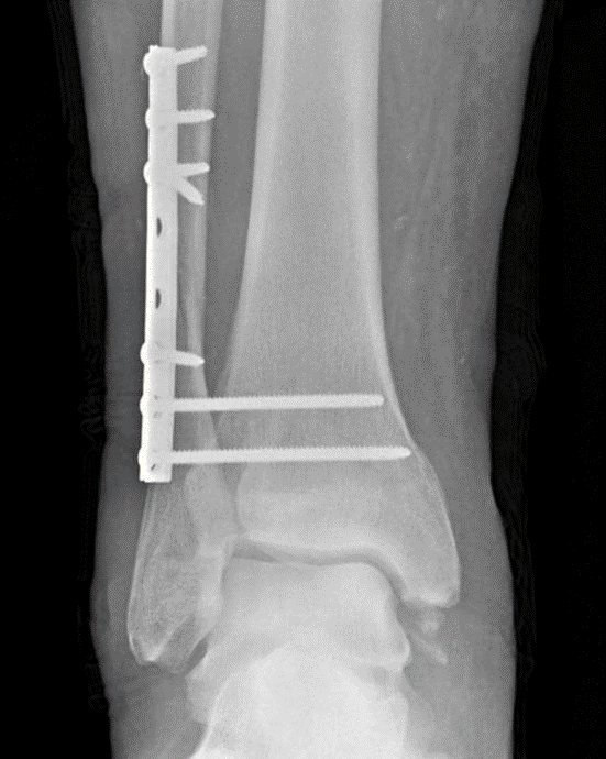
Postoperative radiograph in this patient showing fixation of the fibula and two syndesmotic screws.