Use mouse wheel, arrow keys or left click (with scroll tool selected) to scroll
ui.case.use_touch_gestures
DICOM HelpSource: Local (us-east1-c)
Findings
- Acute, obliquely-oriented distal fibular fracture with inferomedial extension to the level of the tibiotalar joint
- Slight widening of the medial clear space
- Overlying soft tissue swelling
- Remote avulsion fracture at the site of attachment of the ankle joint capsule on the dorsal talus
Diagnosis
Weber type B ankle fracture
Sample Report
Acute, obliquely-oriented distal fibular fracture with inferomedial extension to the level of the tibiotalar joint (Weber B). Overlying soft tissue swelling.
Slight widening of the medial clear space raises concern for deltoid ligament injury.
Remote avulsion fracture at the site of attachment of the ankle joint capsule on the dorsal talus.
Discussion
- While more complex ankle fracture classification systems exist, it is important to at least be familiar with the Weber classification:
- Weber type A: lateral malleolar fracture below the level of the talar dome – these are typically stable and managed conservatively
- Weber type B: lateral malleolar fracture with fracture line exiting medially at the level of the talar dome – these are stable if the medial structures are intact: look for widening of the medial clear space (indicating deltoid ligament injury) and widening of the distal tibiofibular syndesmosis
- Weber type C: lateral malleolar fracture above the level of the talar dome – usually unstable as these are associated with injury to the distal tibiofibular syndesmosis
- Just like the radius and ulna, think about the tibia and fibula as a ring structure. If you see injury in one place (either fracture or joint subluxation) inspect the rest of the ring for additional injuries. If you only see widening of the medial clear space of the ankle mortise, make sure to get tibia/fibula radiographs to look for proximal fibular (Maisonneuve) fracture
Annotated Images & Illustrations
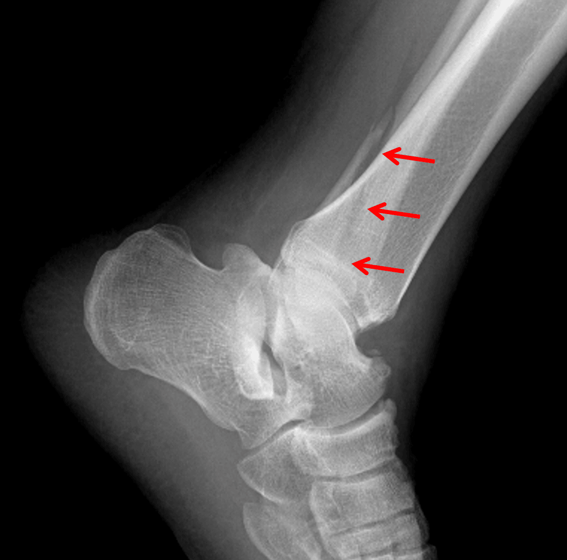
Red arrows: Weber B distal fibular fracture, which is actually best seen on the lateral view in this patient.
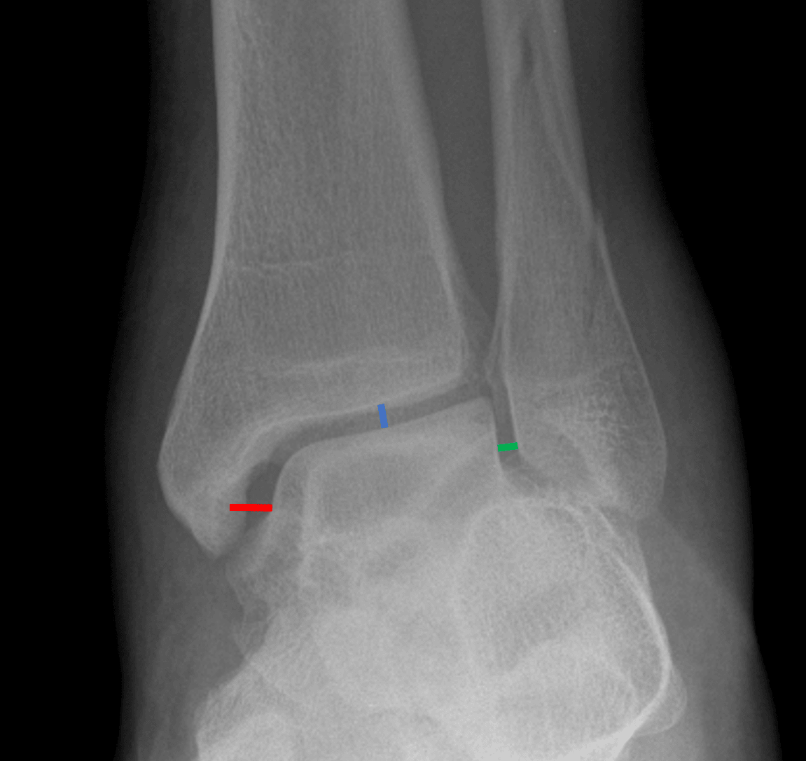
Medial clear space widening. The medial clear space (red line) should not be wider than the other aspects of the ankle mortise (blue and green lines).