Use mouse wheel, arrow keys or left click (with scroll tool selected) to scroll
ui.case.use_touch_gestures
DICOM HelpSource: Local (us-east1-c)
1/189
Keyboard shortcuts (Alt+K)
Demographics:
37 years old, Female
Indication:
Right lower quadrant pain
Findings
Lower chest
- Mild dependent atelectasis
Abdomen/Pelvis
- Dilated retrocecal appendix measuring up to 19 mm in diameter with diffuse wall thickening and multiple appendicoliths measuring up to 12 mm in the base
- No areas of mural hypoenhancement or discontinuity
- Periappendiceal fat stranding without fluid collection or free air
- Several enlarged ileocolic lymph nodes, likely reactive
- IUD in the uterine fundus
MSK
- No acute findings
Diagnosis
Acute appendicitis
Sample Report
Acute suppurative appendicitis. No perforation or abscess.
Discussion
- Acute appendicitis is a common cause for right lower quadrant pain and the diagnosis of exclusion in most patients presenting with this complaint
- Like the gallbladder, the appendix is a blind ending structure that becomes inflamed when something lodges in its neck preventing outflow
- The appendix is considered dilated if it measures greater than 6 mm in diameter, but the upper limit of normal is actually near 10 mm in adults, so if you see gas throughout an appendix that measures more than 6 mm, it is likely normal
- When describing acute appendicitis, make sure to comment on:
- Location - this may change surgical approach, particularly if the appendix is retrocecal
- Presence of appendicoliths - surgeons will try to make sure these are removed and accounted for
- Wall integrity - look for areas of wall nonenhancement or discontinuity to raise concern for necrotic changes
- Periappendiceal collections - look for abscesses and free air (remember that with perforated appendicitis, there will usually only be a trace amount of pneumoperitoneum)
Stages of Acute Appendicitis
Annotated Images & Illustrations
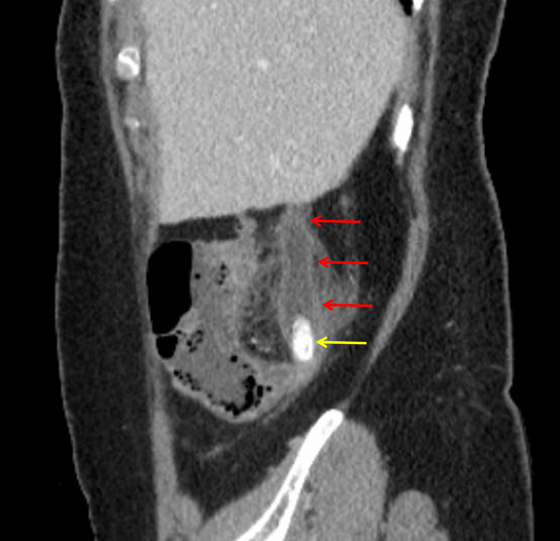
Acute appendicitis: dilated appendix with wall thickening and hyperenhancement (red arrows) and appendicoliths in the base of the appendix (yellow arrow).