1/25
患者信息:
7 岁, 男性
检查指征:
Screening MRI in patient with tuberous sclerosis
影像所见
- Interval enlargement of an enhancing subependymal nodule near the right foramen of Monro measuring 1.5 x 1 cm
- Multiple additional subcentimeter enhancing subependymal nodules
- Multiple T2/FLAIR hyperintense cortical and juxtacortical lesions in the bilateral cerebral hemispheres
- Dilated perivascular spaces in the right greater than left frontal white matter
- No hydrocephalus
- Right maxillary sinus mucous retention cyst. Mucosal thickening of bilateral ethmoid air cells
带注释的图像和插图
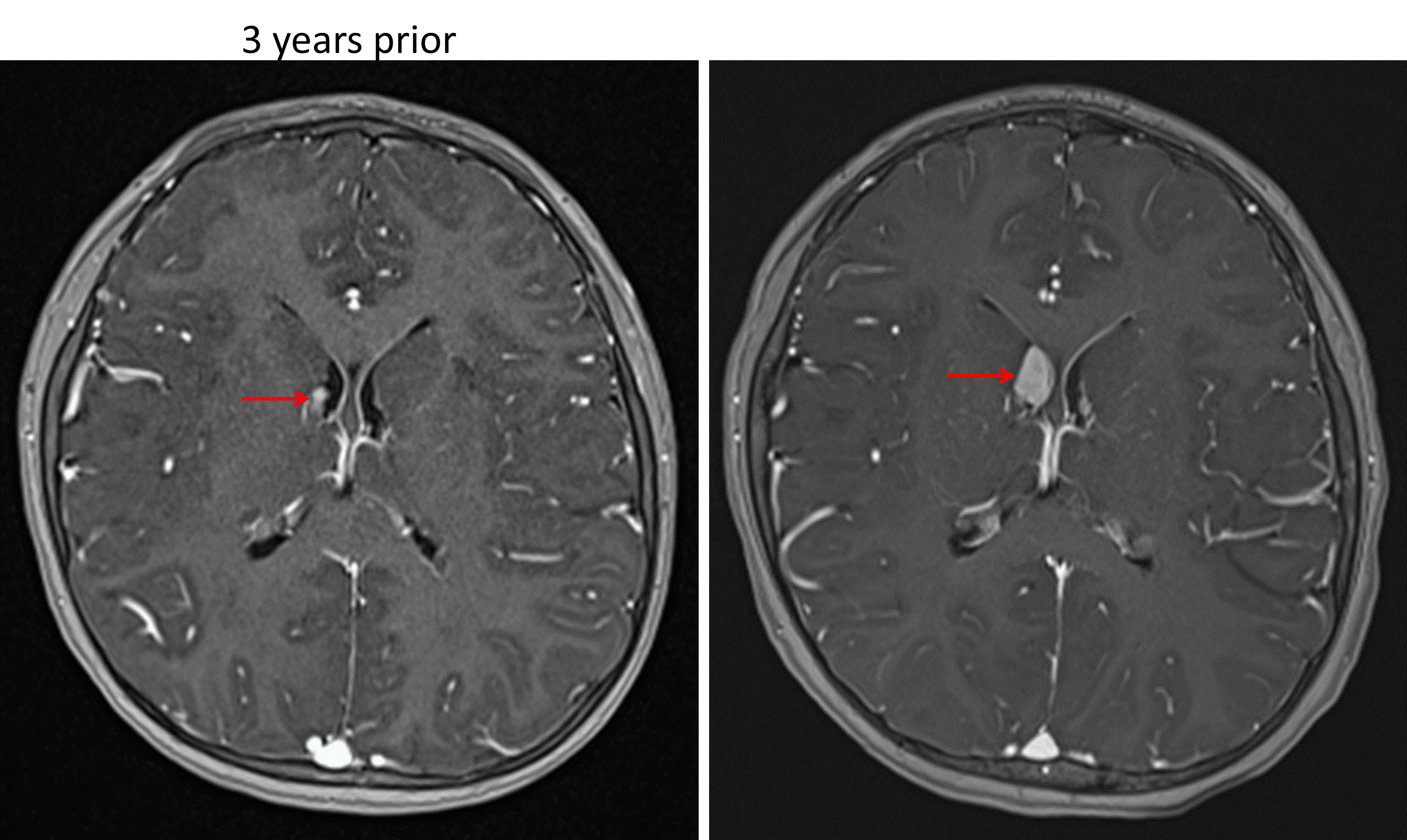
Enhancing subependymal nodule near the right foramen of Monro (red arrows) which demonstrates growth over a 3-year period, concerning for a subependymal giant cell astrocytoma in this patient with tuberous sclerosis.
诊断
Subependymal giant cell astrocytoma (SEGA)
关键影像特征
- SEGAs are enhancing subependymal masses, commonly located near the foramen of Monro
- While SEGAs can be difficult to distinguish from subependymal nodules, growth over time and size exceeding 1 cm favor SEGA
鉴别诊断
- No differential diagnosis in a patient with known tuberous sclerosis
讨论
- Subependymal giant cell astrocytomas (SEGAs) are almost exclusively seen in patients with tuberous sclerosis
- They are thought to arise from subependymal nodules and typically present in later childhood or adolescence
- The primary concern with SEGAs is the development of hydrocephalus due to ventricular outflow obstruction at the foramen of Monro
要点
- In a patient with tuberous sclerosis, an enlarging subependymal nodule exceeding 1 cm in size near the foramen of Monro is highly concerning for a SEGA
- Watch closely for associated obstructive hydrocephalus