患者信息:
31 岁, 男性
检查指征:
踝关节疼痛
诊断
Weber B型踝关节骨折
示例报告
急性斜行腓骨远端骨折,向内下方延伸至胫距关节水平(Weber B型)。覆盖区域软组织肿胀。
内侧间隙轻度增宽提示三角韧带损伤可能。
距骨背侧关节囊附着处陈旧性撕脱骨折。
讨论
- 虽然存在更复杂的踝关节骨折分型系统,但至少应熟悉Weber分型:
- Weber A型:外踝骨折位于距骨穹隆以下——通常稳定,保守治疗
- Weber B型:外踝骨折线内侧端达距骨穹隆水平——如果内侧结构完整则稳定:寻找内侧间隙增宽(提示三角韧带损伤)和胫腓下联合增宽
- Weber C型:外踝骨折位于距骨穹隆以上——通常不稳定,因为与胫腓下联合损伤相关
- 就像桡骨和尺骨一样,将胫骨和腓骨视为环形结构。如果在一处发现损伤(骨折或关节半脱位),需检查环形结构的其余部分是否有额外损伤。如果仅发现踝穴内侧间隙增宽,务必行胫腓骨X线片以寻找腓骨近端(Maisonneuve)骨折
带注释的图像和插图
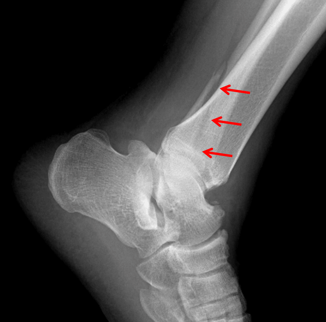
Red arrows: Weber B distal fibular fracture, which is actually best seen on the lateral view in this patient.
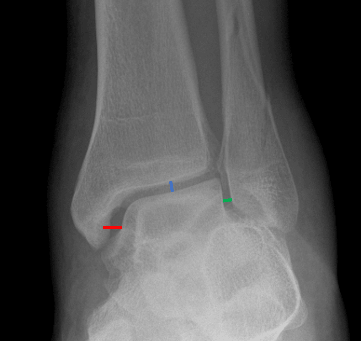
Medial clear space widening. The medial clear space (red line) should not be wider than the other aspects of the ankle mortise (blue and green lines).