Demographics:
12 years old, Male
Indication:
Headache, lethargy
Findings
- T1 hypointense, T2/FLAIR hyperintense, nonenhancing mass measuring 4.6 x 5.5 x 5.8 cm centered in the left thalamus with inferior extension into the left eccentric midbrain, lateral extension into the posterior limb of the left internal capsule, and superior extension into the left frontoparietal periventricular white matter
- Areas of internal restricted diffusion, particularly in the inferior and anterosuperior aspects of the mass
- Associated obstructive hydrocephalus at the level of the posterior third ventricle and cerebral aqueduct with subependymal edema and supratentorial sulcal effacement
- Dilated optic nerve sheaths and expanded Meckel's caves relating to elevated intracranial pressure
- Areas of restricted diffusion in the bilateral parahippocampal gyri which could represent ischemic infarcts, sequelae of recent seizure activity, or less likely additional areas of tumor involvement
Annotated Images & Illustrations
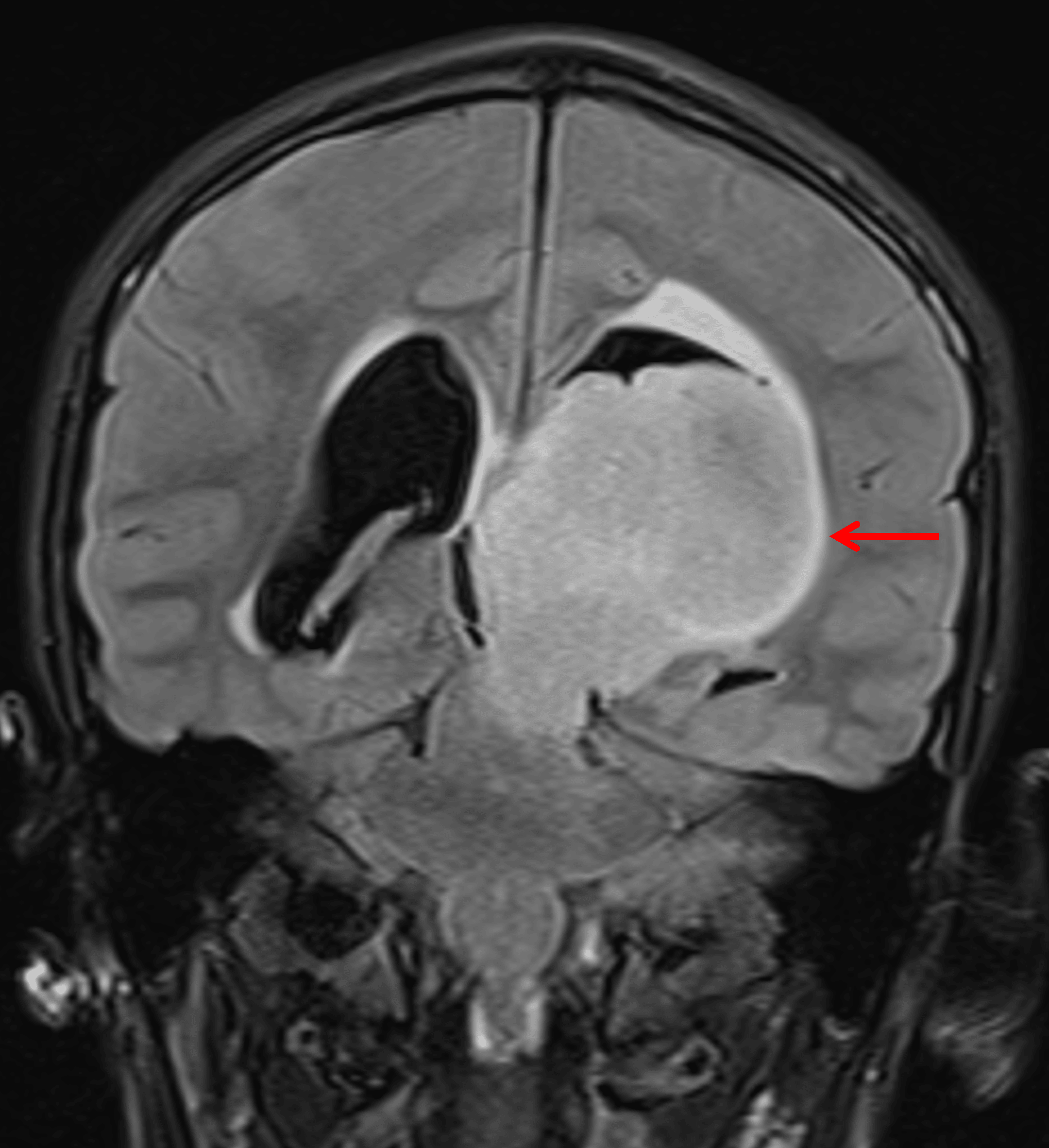
Large, relatively homogeneous T2/FLAIR hyperintense mass centered in the left thalamus (red arrow), consistent with a diffuse midline glioma.
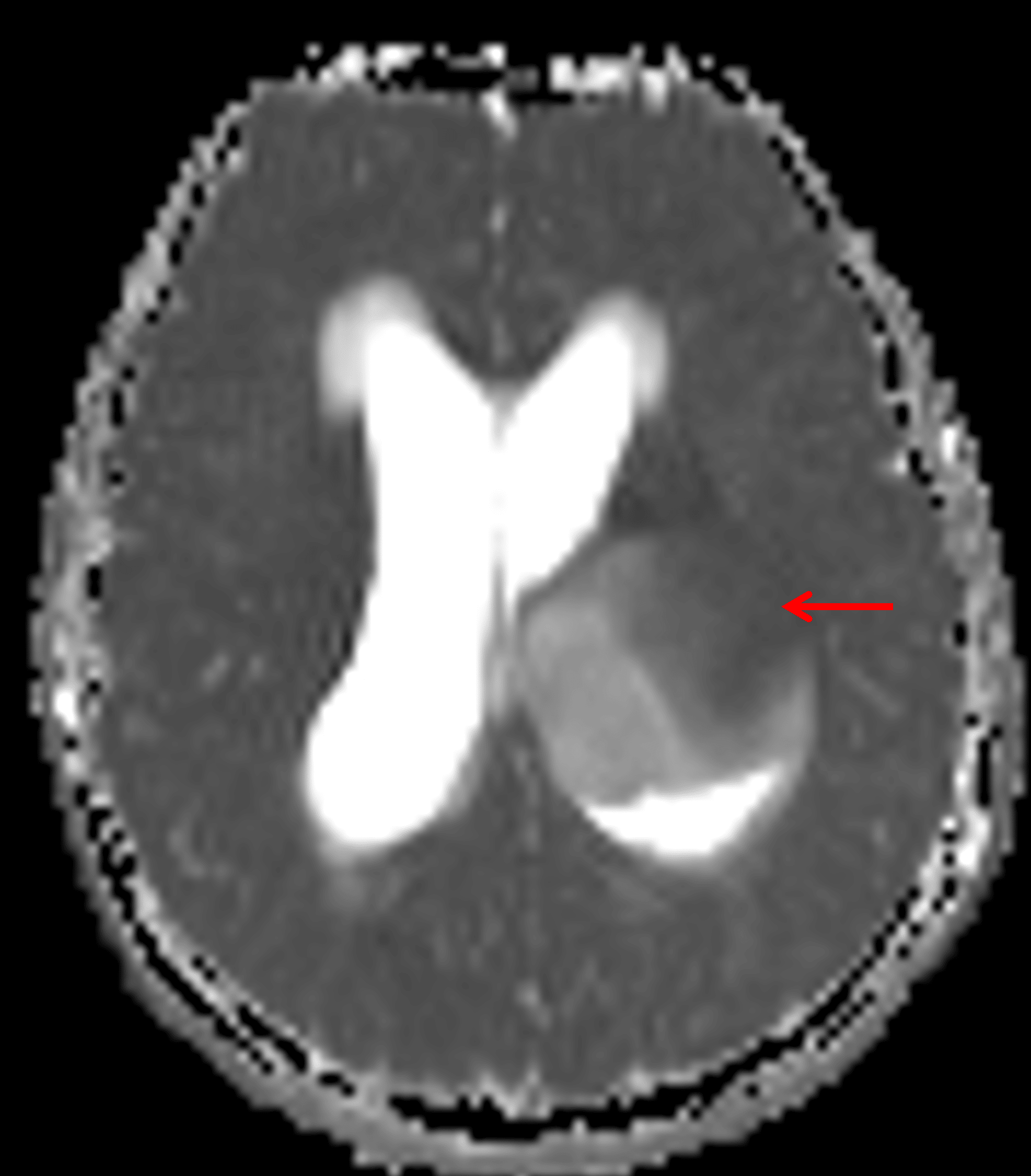
Area of relatively low ADC in the anterosuperior portion of the mass (red arrow), likely correlating with an area of higher cellularity.
Diagnosis
Diffuse midline glioma
Differential Diagnosis
Become a PRO member to unlock the differential diagnosis
Pearls
Become a PRO member to unlock the pearls