Findings
- Extensive plaque-like dural thickening and enhancement with multiple intermixed discrete dural-based masses
- Enhancing lesions in the bilateral internal auditory canals, larger on the right
- Amorphous enhancing lesion along the posterior intraorbital left optic nerve near the orbital apex
- Enhancing soft tissue in the left Meckel's cave extending into the left foramen ovale
- Partially imaged enhancing lesions involving the upper cervical spinal cord
- Prior high left frontoparietal craniotomy with subjacent encephalomalacia in the posterior aspect of the left superior frontal gyrus
- Additional small area of encephalomalacia in the posterior left frontal periventricular white matter with central susceptibility artifact
- The cerebellar tonsils extend up to 8 mm inferior to the foramen magnum with associated mild crowding of the cervicomedullary junction
- Empty, expanded appearance of the sella
- Asymmetric atrophy of the right muscles of mastication
Annotated Images & Illustrations
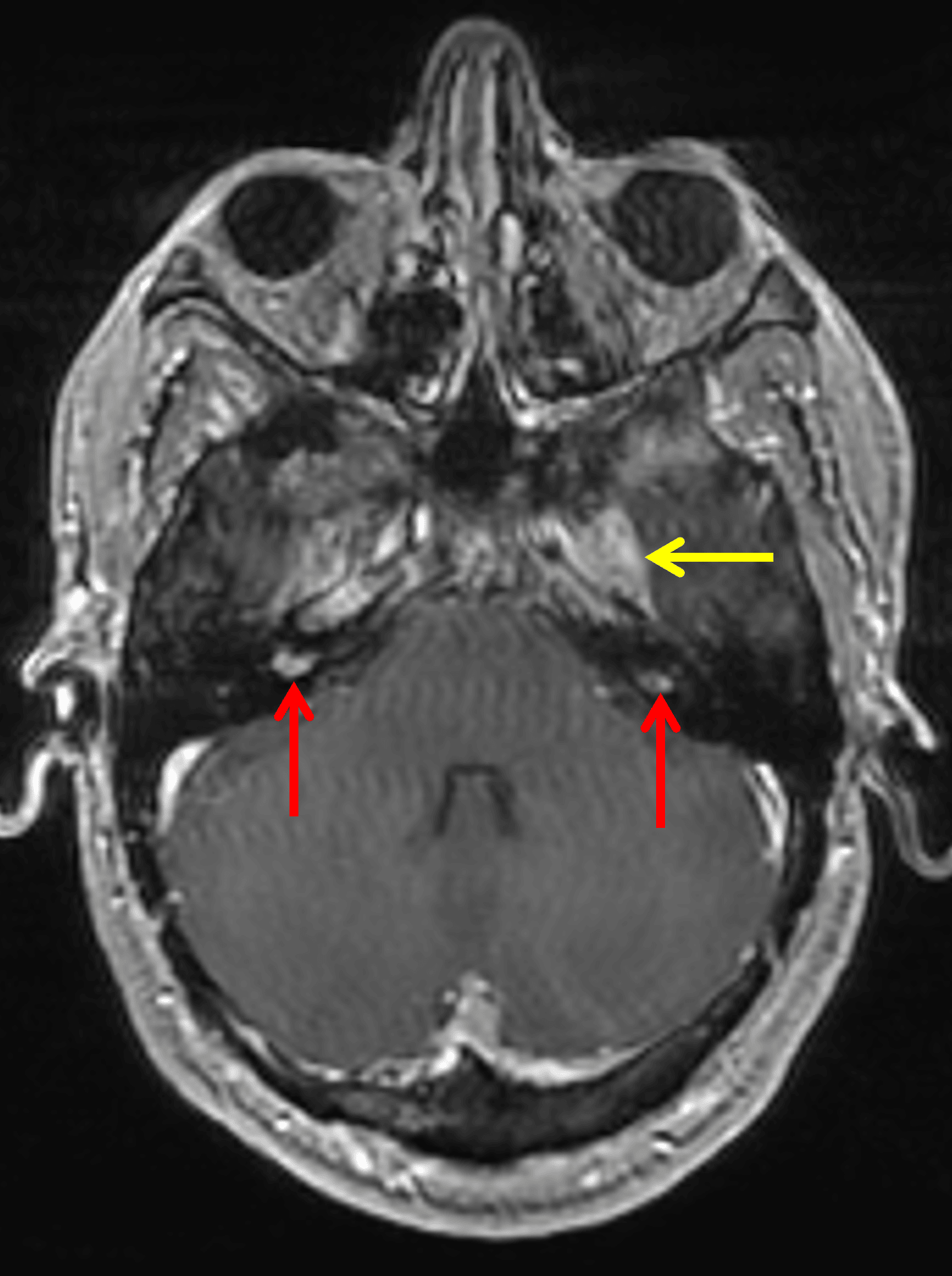
Bilateral vestibular schwannomas (red arrows). Enhancement in the left Meckel's cave extending into the foramen ovale (yellow arrow), also likely a schwannoma.
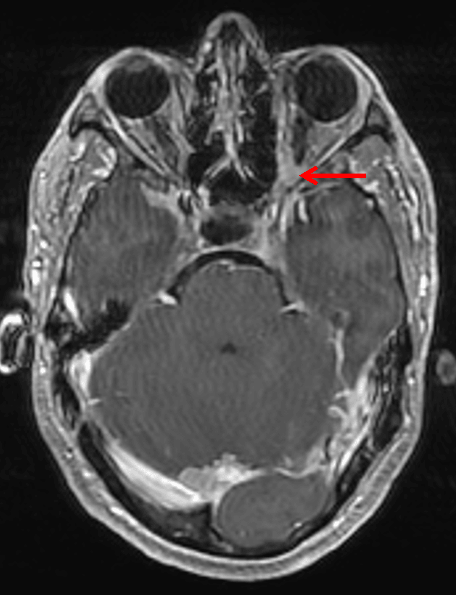
Enhancement in the left orbital apex along the optic nerve (red arrow), likely representing an optic nerve sheath meningioma.
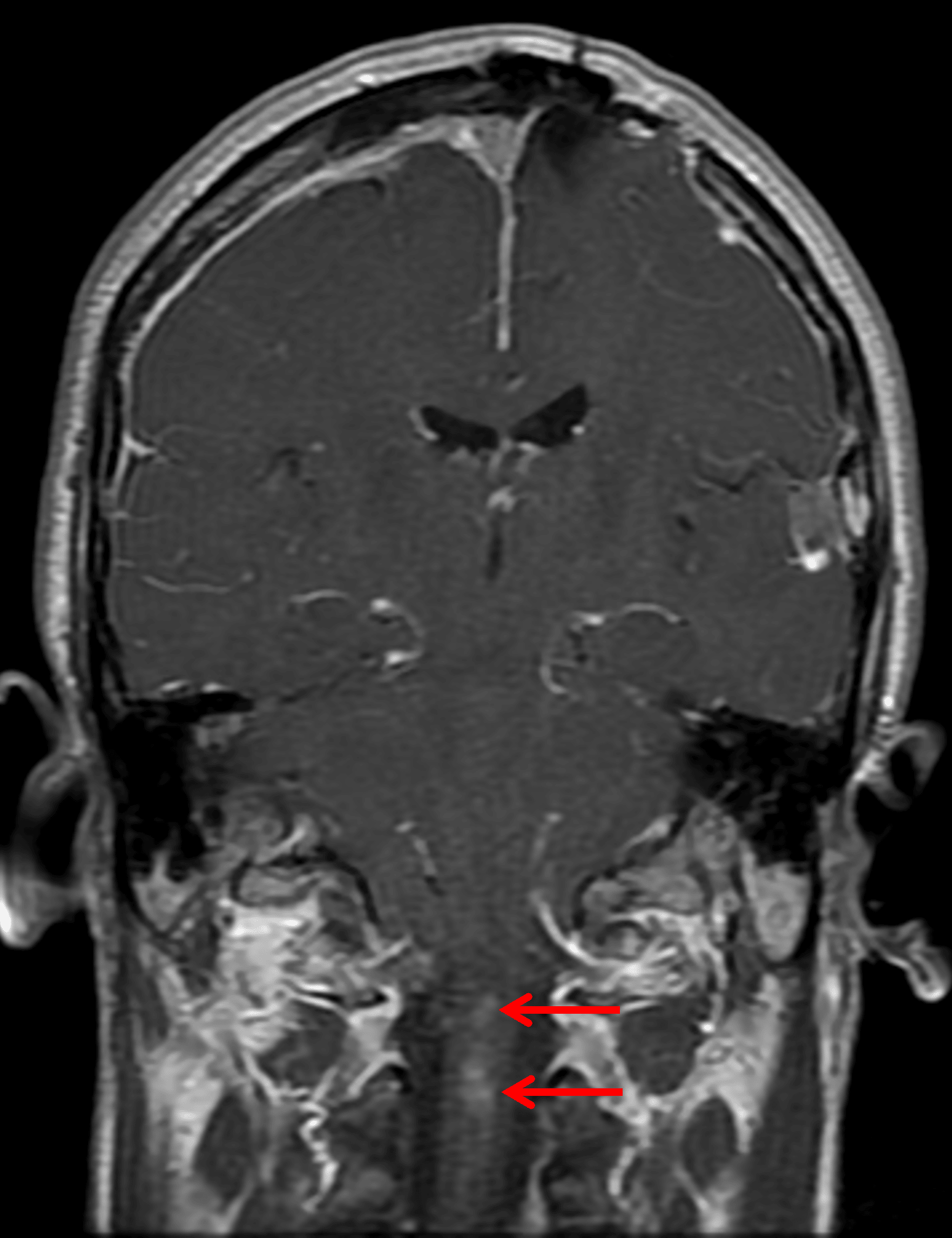
Enhancing intramedullary lesions in the upper cervical spinal cord (red arrows), likely representing ependymomas.
Diagnosis
Meningiomatosis, schwannomas, and ependymomas in neurofibromatosis type 2
Key Imaging Features
Become a PRO member to unlock the key imaging features
Differential Diagnosis
Become a PRO member to unlock the differential diagnosis
Discussion
Pearls
Become a PRO member to unlock the pearls