Demographics:
57 years old, Male
Indication:
Gait ataxia
Findings
- Patchy and confluent areas of T2/FLAIR signal hyperintensity in the bilateral cerebral white matter, advanced for patient age
- Corresponding patchy restricted diffusion
- Corresponding faint linear and nodular areas of enhancement in the bilateral frontal white matter and to a lesser extent in the bilateral parietal white matter
- Intermixed foci of susceptibility artifact
- No substantial intracranial mass effect or evidence of hydrocephalus
Annotated Images & Illustrations
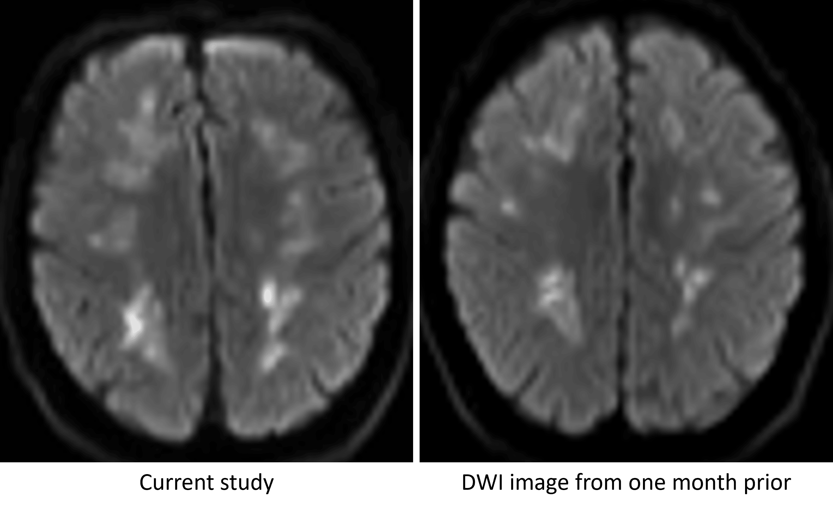
Persistence of restricted diffusion in the cerebral white matter over a one month interval, which is not typical for ischemic infarcts resulting from a single event.
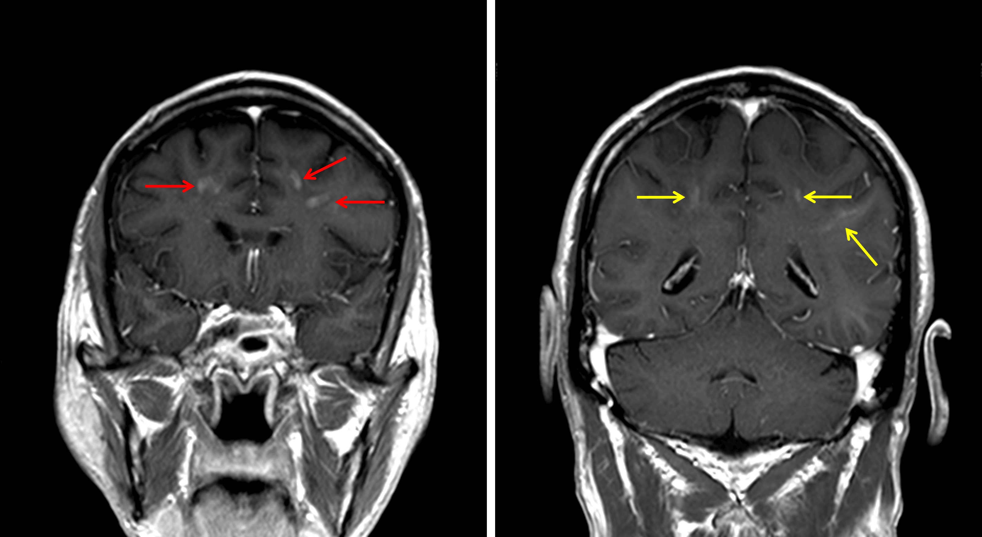
Corresponding areas of enhancement in the frontal lobes (red arrows) and to a lesser extent in the parietal lobes (yellow arrows).
Diagnosis
Intravascular lymphoma
Differential Diagnosis
Become a PRO member to unlock the differential diagnosis
Pearls
Become a PRO member to unlock the pearls