Demographics:
4 years old, Female
Indication:
History of neurofibromatosis type 1
Findings
- Enlargement of the right greater than left prechiasmatic optic nerves and optic chiasm, likely representing optic pathway glioma
- Patchy, minimally expansile T2/FLAIR signal hyperintensity involving the anterior commissure, left forniceal column, bilateral deep gray structures, brainstem, and left greater than right cerebellar white matter without corresponding restricted diffusion or enhancement, which may represent focal areas of signal intensity (FASI) and/or infiltrative glioma
- Left persistent trigeminal artery
- Developmental venous anomaly involving the right frontal operculum
Annotated Images & Illustrations
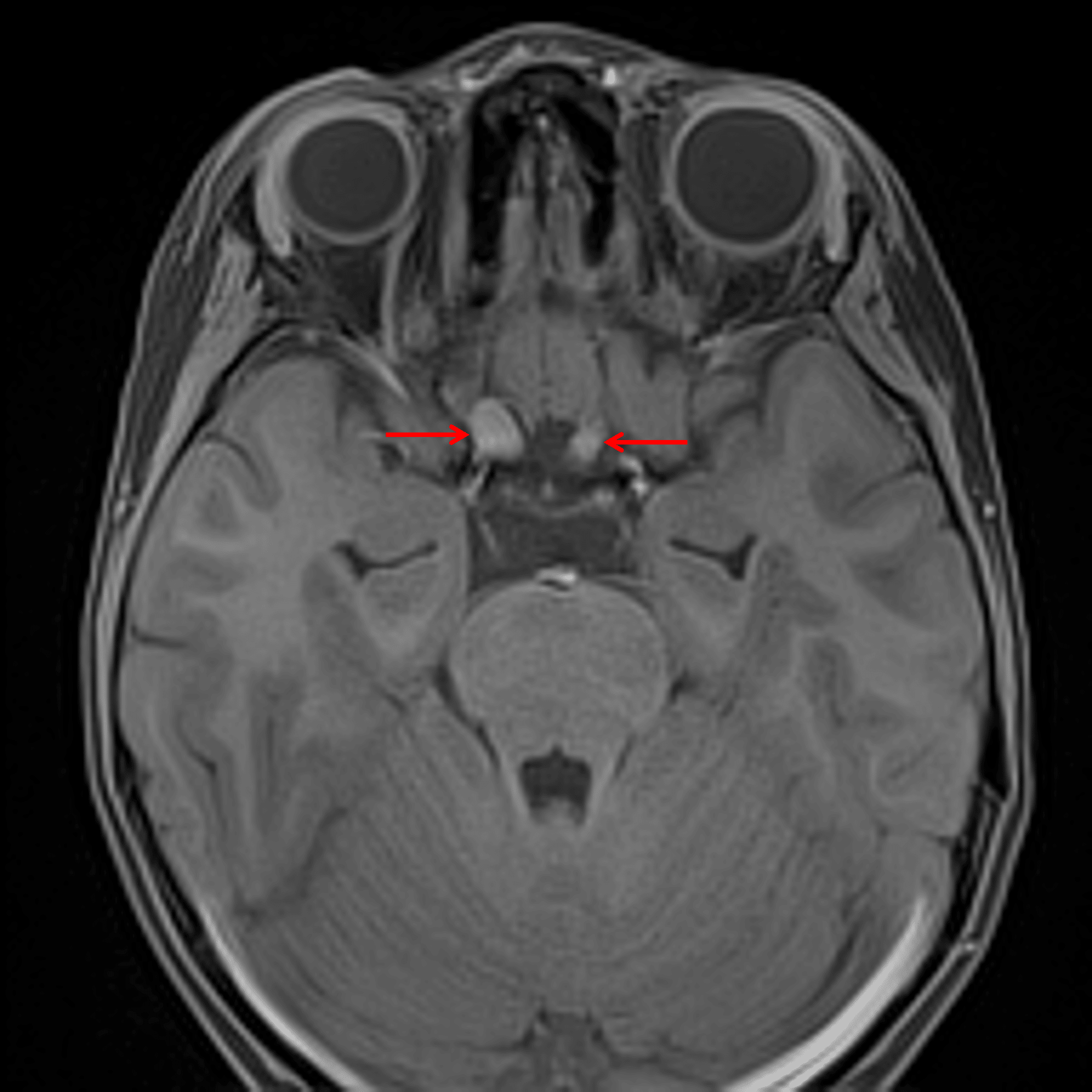
Enlargement of the right greater than left prechiasmatic optic nerves (red arrows), consistent with optic pathway glioma in this patient with NF-1.
Diagnosis
Optic pathway glioma
Key Imaging Features
Become a PRO member to unlock the key imaging features
Differential Diagnosis
Become a PRO member to unlock the differential diagnosis
Discussion
Pearls
Become a PRO member to unlock the pearls