Demographics:
64 years old, Female
Indication:
Headache
Findings
CT
- Calcified suprasellar mass
MRI
- T1/T2/FLAIR isointense, diffusely enhancing suprasellar mass measuring 1.8 x 1.4 x 1.7 cm
- Predominantly peripheral corresponding susceptibility artifact corresponding with calcification seen on CT
- The mass is centered on the pituitary stalk and is seen separate from the pituitary gland
- The mass contacts the optic tracts and posterior aspect of the optic chiasm
Annotated Images & Illustrations
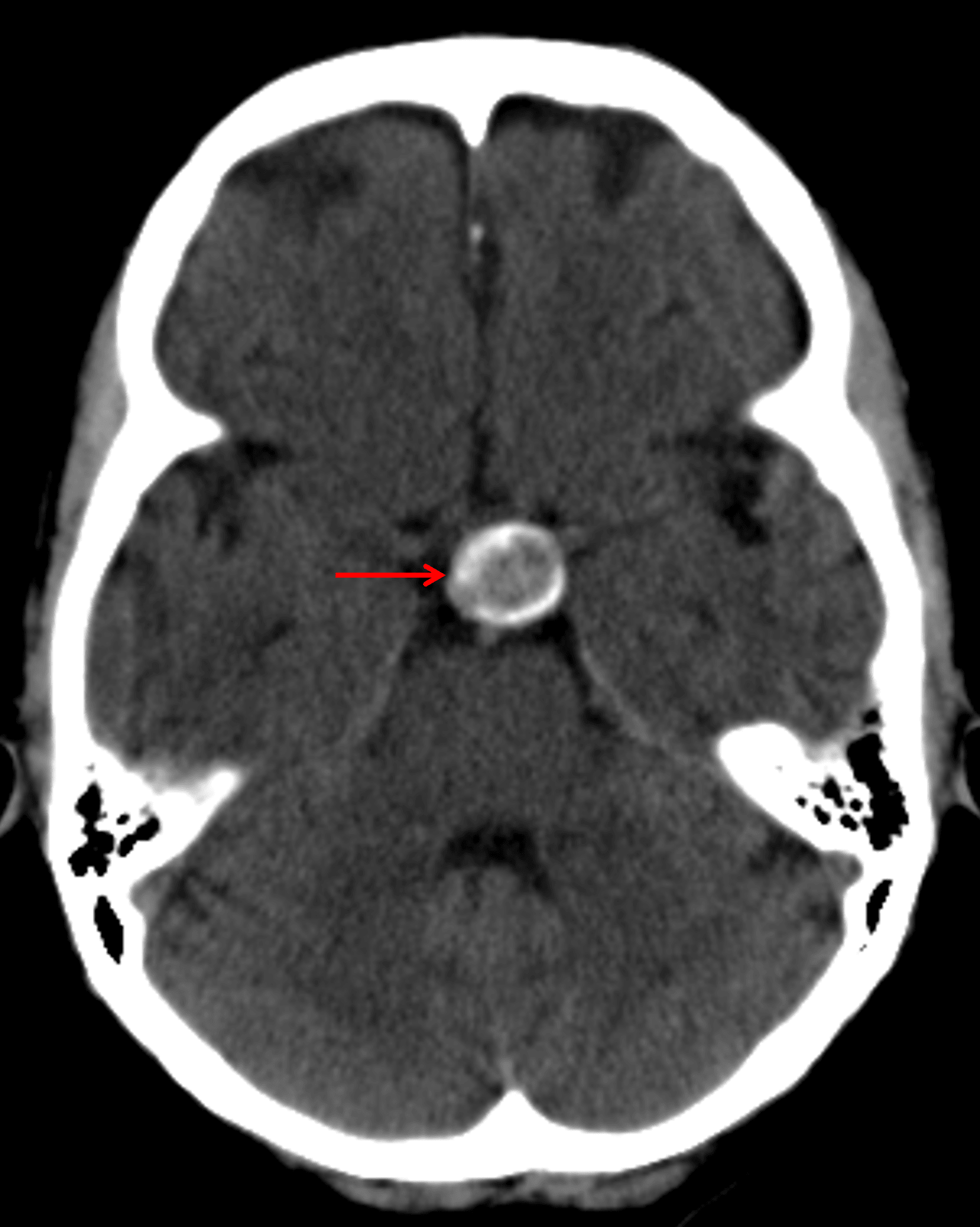
Calcified suprasellar mass (red arrow).
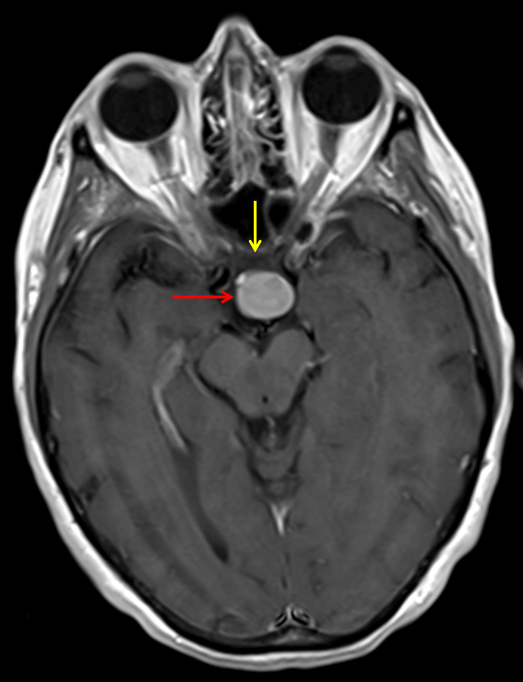
Diffuse corresponding enhancement (red arrow). The mass contacts the posterior aspect of the optic chiasm (yellow arrow).
Diagnosis
Papillary craniopharyngioma
Differential Diagnosis
Become a PRO member to unlock the differential diagnosis
Pearls
Become a PRO member to unlock the pearls