マウスホイール、矢印キー、または左クリック(スクロールツール使用時)でスクロールしてください。
ui.case.use_touch_gestures
DICOMヘルプSource: Local (us-east1-c)


症例 #1
所見
- 左尾状核視床溝のエコー輝度の高い物質および左側脳室後角に沈殿するエコー輝度の高い物質
- 脳室拡大なし
- 異常な脳実質エコー輝度または軸外液貯留なし
- 未熟な脳溝パターン
診断
グレード2胚層板出血
サンプルレポート
左尾状核視床溝に及び左側脳室後角に沈殿する左胚層板出血で、水頭症を伴わない(グレード2)。
異常な脳実質エコー輝度または軸外液貯留なし。
未熟な脳溝パターン。
考察
- 胚層板は高度に血管化した上衣下構造で、周産期ストレスの結果として出血のリスクがある
- 胚層板は妊娠第3期に退縮する。32週までに尾状核視床溝に限局し、36週までにほぼ消失する。したがって、36週以降に出生した児では胚層板出血のリスクは実質的にゼロであり、逆に在胎32週未満で出生した児ではリスクが大幅に増加する
- 症状が非特異的である可能性があるため、在胎30週未満で出生した児にはスクリーニング神経超音波検査が推奨される
- 胚層板出血を探す際、脈絡叢(残念ながら出血と同様にエコー輝度が高い)は尾状核視床溝より前方には伸展しないはずであるため、前角のエコー輝度の高い物質は出血の可能性が高いことを覚えておくこと
- 以下は胚層板出血の4段階分類である:
胚層板出血
- 水頭症 - 以下は超音波での概算正常計測基準である(Brouwer et al.より)。これらの計測例は以下の注釈付き画像で示されている
- 脳室指数(冠状断で計測した前角外側壁から大脳鎌までの距離):25〜30週で < 10 mm、40週で < 15 mm
- 前角幅(冠状断で計測した前角の最大対角線幅):25〜30週で < 3 mm、40週で < 4 mm
- 視床後頭距離(矢状断で計測した視床後縁から後角後縁までの距離):25〜30週で < 20 mm、40週で < 25 mm
- 脳が成熟するにつれ、脳溝はより深く、より多くなる
- 患児の月齢に対する正常な脳溝パターンを認識することは、滑脳症などの皮質発達障害の早期診断に有用である
- 大きな脳溝/裂(例:シルビウス裂や頭頂後頭溝)は発達の早期に明瞭になるが、小さな脳溝は妊娠第3期の後期まで超音波で明瞭にならないことがある
- この患児と在胎35週および42週で撮影された患児の脳溝の違いについては、以下の注釈付き画像を参照すること
注釈付き画像とイラスト
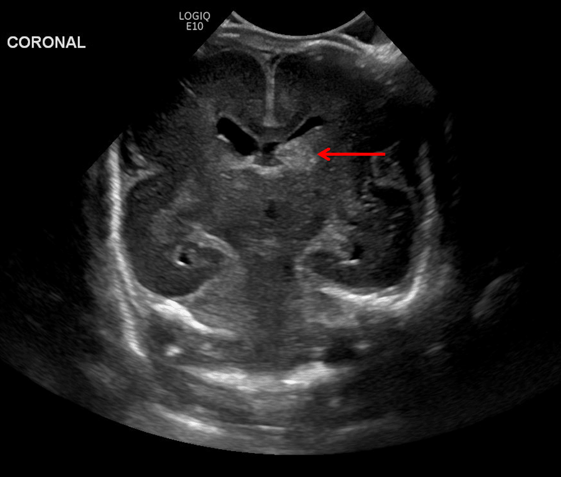
Rounded echogenic area in the left caudothalamic groove (red arrow) consistent with germinal matrix hemorrhage.
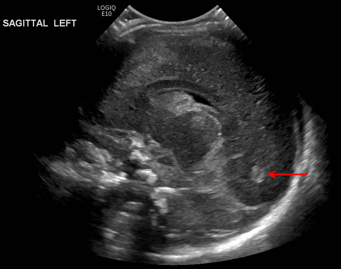
Echogenic material layering in the occipital horn of the left lateral ventricle (red arrow) consistent with intraventricular hemorrhage.
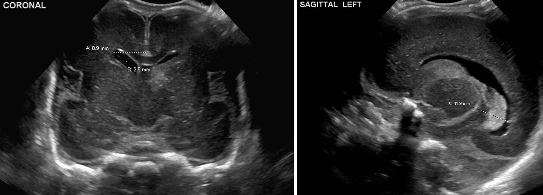
Example measurements of the ventricular index (A), anterior horn width (B), and thalamo-occipital distance (C).
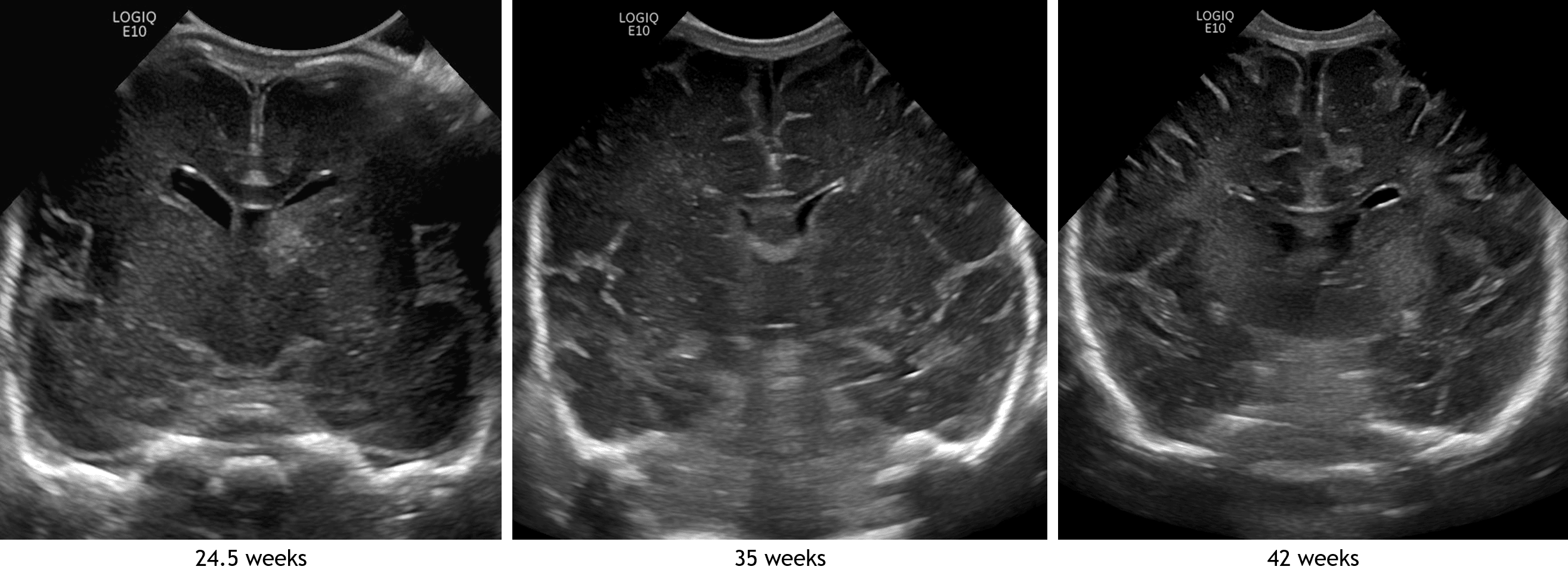
Coronal image in this patient compared with images from different patients imaged at 35 and 42 weeks. Notice how the echogenic sulci become deeper and more numerous with increasing patient age.
参考文献