1/36
Keyboard shortcuts (Alt+K)
Demographics:
71 years old, Male
Indication:
Altered mental status
Findings
- Hypoattenuation and loss of gray-white differentiation involving the left paramedian frontal and parietal lobes
- Remote left basal ganglia lacunar infarct
- Sequelae of chronic small vessel disease
Diagnosis
Left ACA territory infarct
Sample Report
Hypoattenuation and loss of gray-white differentiation involving the left paramedian frontal and parietal lobes consistent with acute/early subacute left ACA territory infarct. No significant associated mass effect or evidence of hemorrhagic conversion.
Remote left basal ganglia lacunar infarct.
Sequelae of chronic small vessel disease.
Discussion
- ACA territory infarcts are much less common than MCA or PCA territory infarcts, in part because if the A1 segment is occluded, the other ACA is typically able to supply both ACA territories via the anterior communicating artery
- While ACA infarcts are most often embolic, severe midline shift can compress the ACAs leading to ischemia
Annotated Images & Illustrations
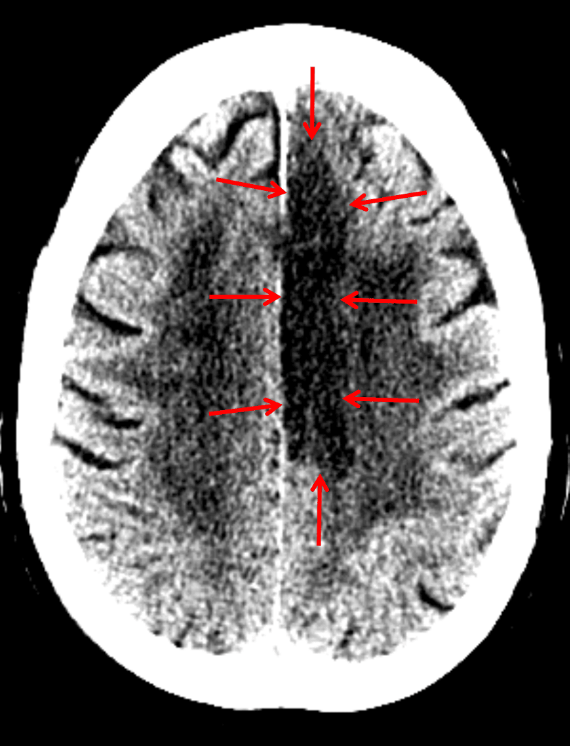
Red arrows: ACA territory infarct
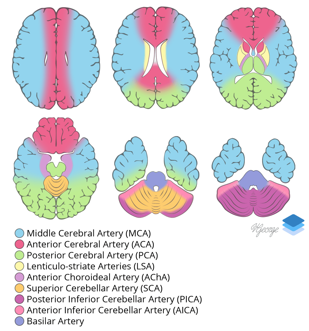
Cross-sectional arterial territories of the brain.
Related Video
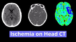
Ischemia on Head CT
YouTube