Findings
- Confluent low attenuation with loss of gray-white differentiation in the left cerebral hemisphere involving the ACA and MCA territories
- Asymmetrically hyperattenuating left MCA and ACA
- Left hemispheric sulcal effacement with slight left to right midline shift and slight effacement of the left lateral ventricle
Diagnosis
Acute left ACA and MCA territory infarct
Sample Report
Diffuse, confluent low attenuation with loss of gray-white differentiation in the left cerebral hemisphere involving the ACA and MCA territories concerning for acute/early subacute infarct. Asymmetrically hyperattenuating appearance of the left MCA and ACA is concerning for thrombosis. Recommend head CTA and brain MRI for further evaluation.
No evidence of hemorrhagic transformation.
Associated cytotoxic edema with resultant left hemispheric sulcal effacement, 3 mm left to right midline shift, and mild narrowing of the left lateral ventricle. The basal cisterns are patent without evidence of herniation.
Discussion
- Remember to always look at the brain on narrow ("stroke") windows
- Dense vessel signs are immediate findings in strokes and are apparent hours before cytotoxic edema becomes apparent on CT. While dense vessel signs are seen in a minority of cases, it is important to look for them because they may be the only clue that the patient is having an acute stroke. However, many technical factors (e.g. beam hardening artifact) can create a pseudo-dense vessel appearance, so it's important to consider the clinical scenario before questioning a dense vessel sign.
Annotated Images & Illustrations
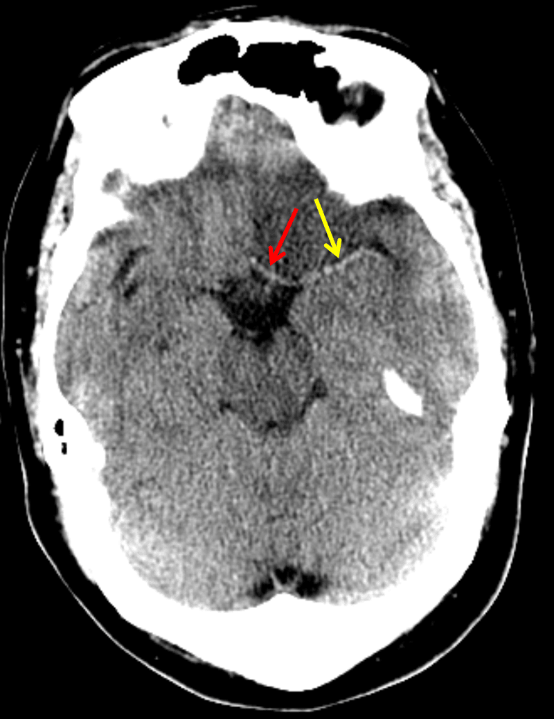
Asymmetrically hyperattenuating appearance of the proximal left ACA (red arrow) and MCA (yellow arrow) concerning for thrombosis
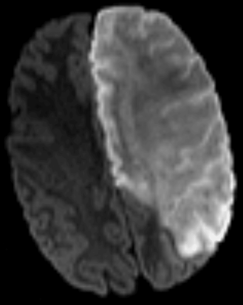
Representative diffusion-weighted MR image from this patient
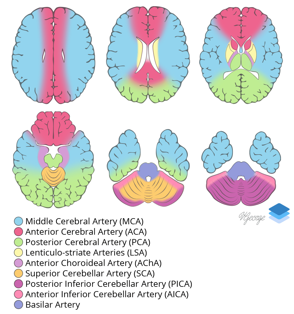
Cross-sectional arterial territories of the brain.
Related Video
Ischemia on Head CT
YouTube