Keyboard shortcuts (Alt+K)
Demographics:
59 years old, Male
Indication:
Motorcycle accident
Findings
- Diastasis of the pubic symphysis measuring up to 3 cm
- Mild widening of the right sacroiliac joint
- Mild degenerative changes of the hips, sacroiliac joints, and of the partially visualized lower lumbar spine
- Arterial and vas deferens calcifications
Diagnosis
Unstable pelvic ring injury (APC II)
Sample Report
Unstable pelvic ring injury with diastasis of the pubic symphysis measuring up to 3 cm and mild widening of the right sacroiliac joint.
Mild degenerative changes of the hips, sacroiliac joints, and of the partially visualized lower lumbar spine.
Arterial and vas deferens calcifications.
Discussion
- It is important to be able to recognize unstable pelvic injuries because this impacts immediate patient management (pelvic binders are typically placed before the patient even goes for CT imaging)
- The Young Burgess classification is the most commonly used system:
- Anterior-posterior compression (APC) - the pubic symphysis is the weak link, so it is affected first
- APC I: widening of the pubic symphysis < 2.5 cm
- Stable
- APC II: widening of the pubic symphysis > 2.5 cm, anterior sacroiliac joint widening
- Vertically stable, rotationally unstable
- APC III: widening of the pubic symphysis > 5 cm, widening of anterior and posterior sacroiliacs joints
- Globally unstable
- APC I: widening of the pubic symphysis < 2.5 cm
- Lateral compression - the obturator ring is the weak link, so it is affected first
- LC I: obturator ring fractures and sacral compression fractures
- Stable
- LC II: LC I + iliac crescent fracture
- Vertically stable, rotationally unstable
- LC III: LC I/II with a contralateral APC injury
- Globally unstable
- LC I: obturator ring fractures and sacral compression fractures
- Vertical shear - Vertical hemipelvis translation with offset of the pubic symphysis and sacroiliac joint
- Globally unstable
- Anterior-posterior compression (APC) - the pubic symphysis is the weak link, so it is affected first
Annotated Images & Illustrations
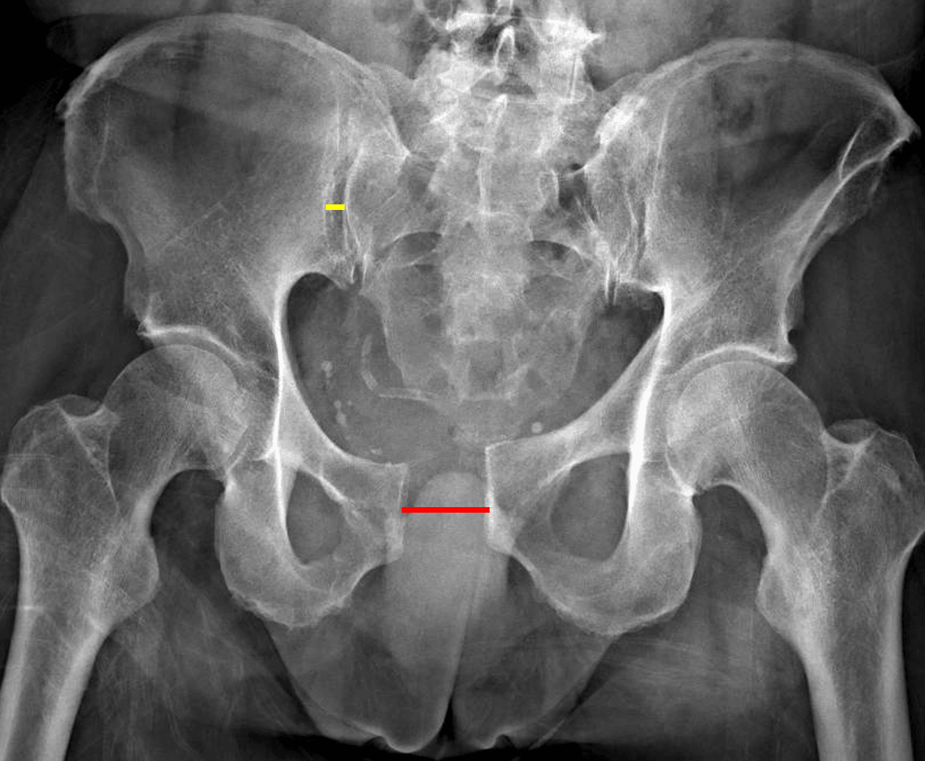
APC II unstable pelvic ring injury with widening of the pubic symphysis (red line) and anterior right sacroiliac joint (yellow line).
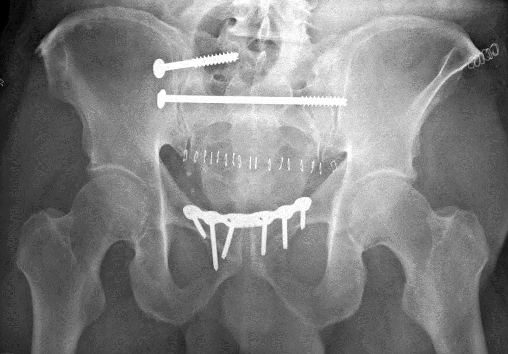
Postoperative radiograph in this patient.