Use mouse wheel, arrow keys or left click (with scroll tool selected) to scroll
ui.case.use_touch_gestures
DICOM HelpSource: Local (us-east1-c)
Keyboard shortcuts (Alt+K)
Demographics:
46 years old, Male
Indication:
Diffuse abdominal pain
Findings
- Multiple dilated loops of small bowel throughout the abdomen measuring up to 5 cm in diameter with multiple air-fluid levels on upright imaging
- Paucity of colonic and rectal gas
- No evidence of pneumoperitoneum on upright images
- Enlarged cardiopericardial silhouette
Diagnosis
Small bowel obstruction
Sample Report
Findings are concerning for high-grade distal small bowel obstruction. No evidence of pneumoperitoneum. Consider CT for further evaluation.
Discussion
- Although small bowel obstruction is becoming more and more a CT diagnosis, it is important to be familiar with the radiographic findings. Some important findings to look for include:
- Gaseous distension of small bowel with bowel loops measuring > 3 cm in diameter
- Air fluid levels measuring at least 2.5 cm in width, at different levels from each other on upright or decubitus imaging
- “String of beads” appearance of tiny locules of gas trapped within predominantly fluid-filled bowel loops
- “Stepladder” appearance of dilated small bowel loops stacked upon each other in coiled array (like a tightly coiled sausage)
- Disproportionate small bowel dilation with paucity of distal/colonic gas
- A proximal small bowel obstruction may only present with gastric distension and/or a focally dilated bowel loop in the upper abdomen
- If the bowel loops are all fluid-filled, the appearance may instead be that of an essentially gasless abdomen
- Make sure to always look for signs of complications such as pneumoperitoneum, pneumatosis, and portal venous gas
Annotated Images & Illustrations
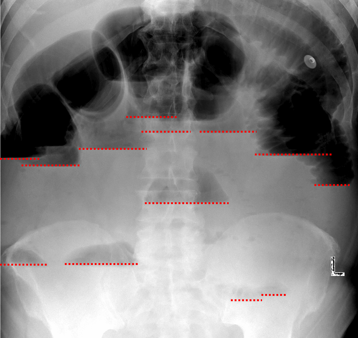
Small bowel obstruction. Red dotted lines highlight many air-fluid levels at different levels in the abdomen on upright imaging.