


患者信息:
8 岁, 男性
检查指征:
右下腹疼痛
病例 #1
影像所见
- 右下腹管状、盲端结构,直径9 mm,不可压缩,伴轻度管壁增厚和轻度壁内血流增多
- 其浅面可见2 x 2.5 cm卵圆形低回声结构,与邻近肠管分界清楚,内部无血流信号
- 右下腹周围炎性改变
诊断
阑尾炎伴阑尾周围脓肿
示例报告
阑尾扩张,直径9 mm,不可压缩,伴轻度管壁增厚和轻度壁内血流增多,考虑急性阑尾炎。
其浅面可见2 x 2.5 cm卵圆形低回声结构,与邻近肠管分界清楚,内部无血流信号,考虑阑尾周围脓肿。
讨论
- 记住超声是儿童和消瘦年轻成人良好的初始检查
- 评估阑尾炎患者时,务必寻找并发症征象,如阑尾周围脓肿和穿孔证据(包括邻近游离气体),因为这些可能影响干预的性质和时机
- 脓肿内部不应有血流信号,但周围常见血流增多
带注释的图像和插图
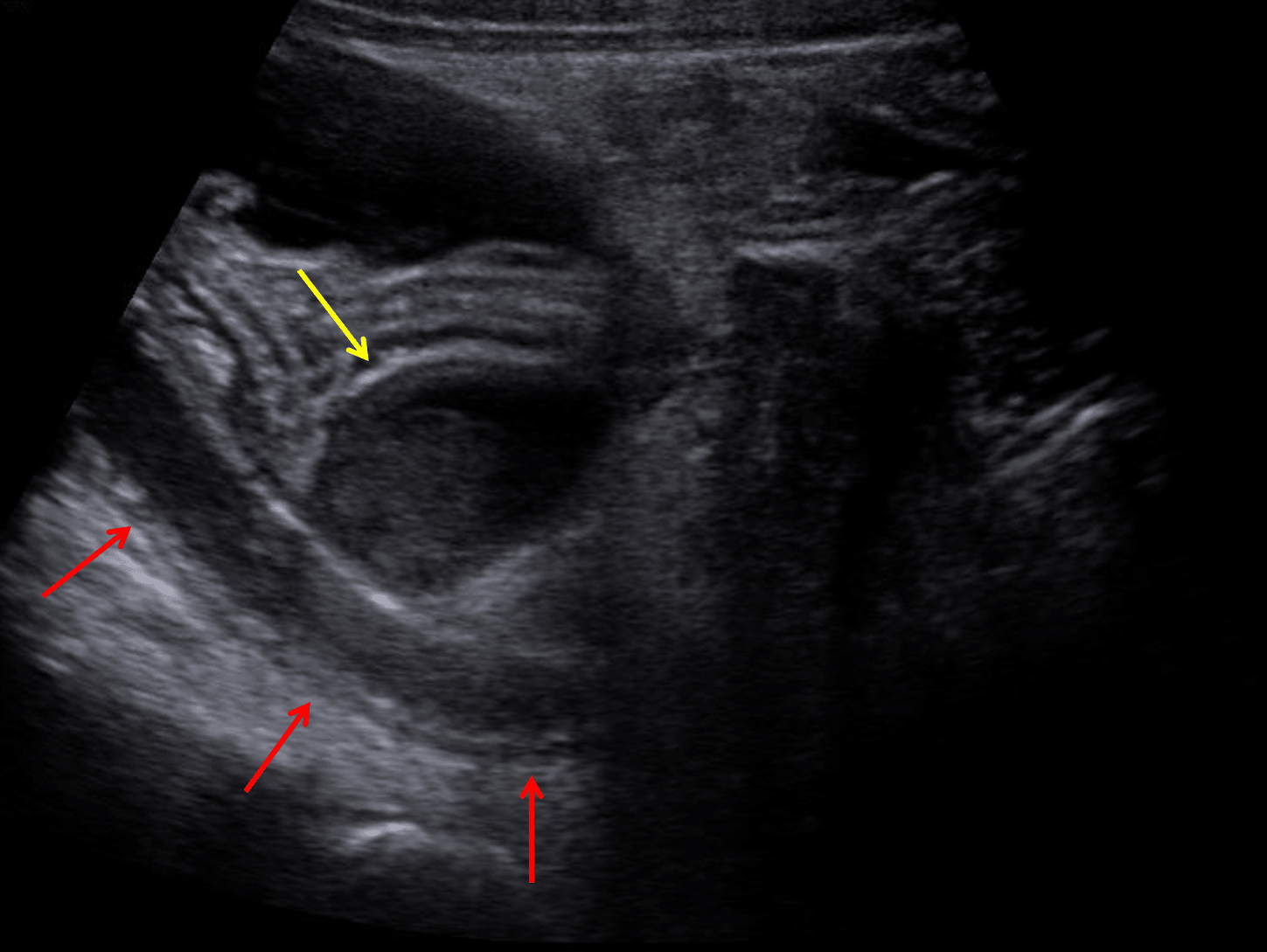
Dilated appendix with mild wall thickening (red arrows). Superficial to the appendix is a hypoechoic structure (yellow arrow), which did not show internal vascularity on color Doppler analysis and is concerning for an abscess.