마우스 휠, 화살표 키 또는 (스크롤 도구 선택 시) 왼쪽 클릭을 사용하여 스크롤하십시오.
ui.case.use_touch_gestures
DICOM 도움말Source: Local (us-east1-c)
1/30
환자 정보:
38 세, 남성
적응증:
Seizure
소견
- T1 hypointense, T2 hyperintense expansile mass extending to the cortex in the left superior frontal gyrus
- Mixed hyperintense and isointense internal FLAIR signal
- No corresponding enhancement, restricted diffusion, or susceptibility artifact
- Mild corresponding local mass effect without midline shift or evidence of herniation or hydrocephalus
주석이 있는 이미지 및 일러스트
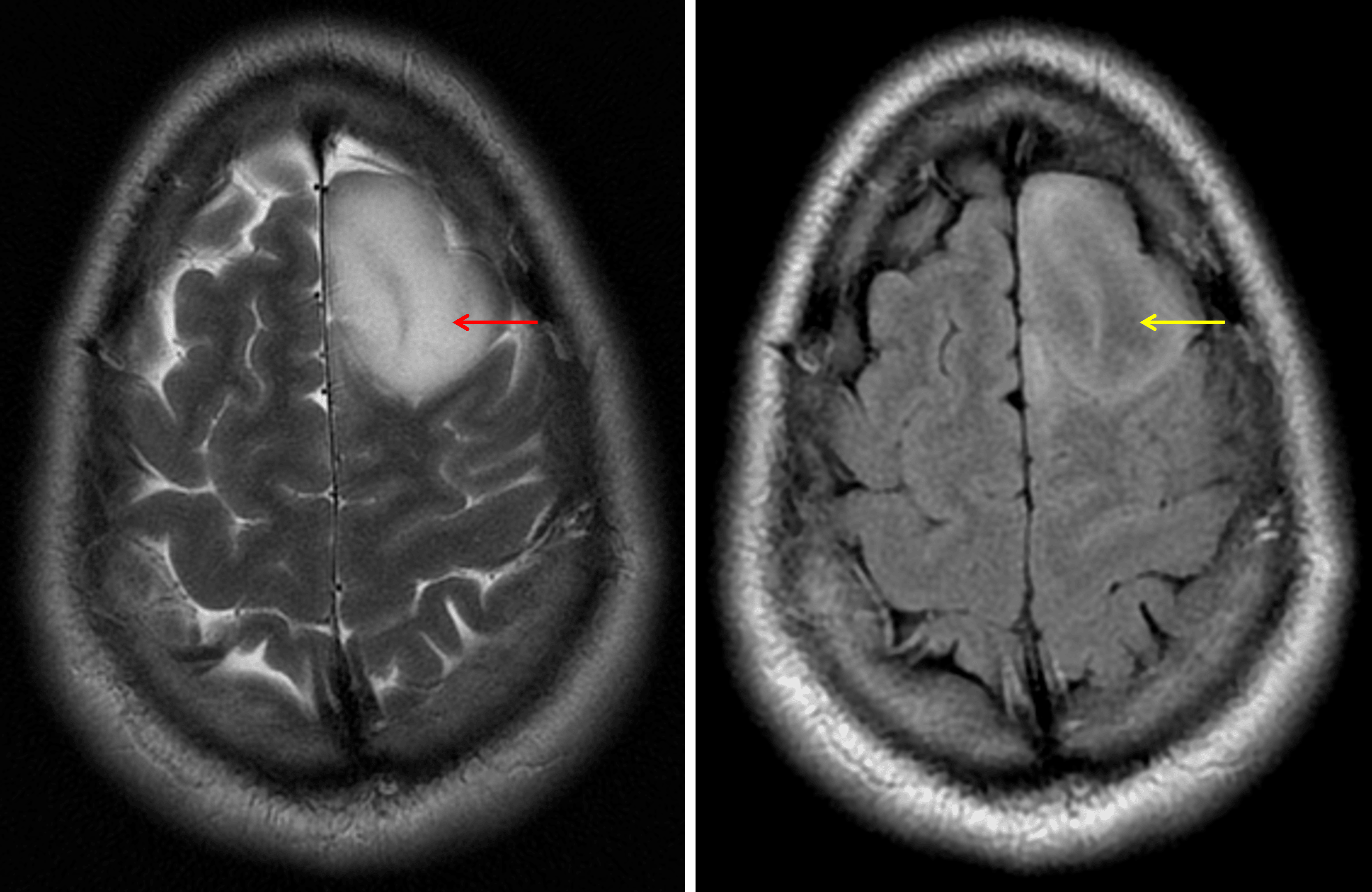
The T2-FLAIR mismatch sign: the lesion demonstrates internal T2 signal hyperintensity (red arrow) contrasted to relatively hypointense FLAIR signal (yellow arrow).
진단
IDH-mutant astrocytoma
감별진단
- Dysembryoplastic neuroepithelial tumor (DNET) - This case is actually a very good look for a DNET, specifically bubbly internal T2 signal hyperintensity, signal suppression on FLAIR, and lack of contrast enhancement. DNETs are often smaller than the tumor in this case
- Ganglioglioma - These tumors are also often peripheral, but commonly enhance and have associated calcification. These tumors more frequently involve the temporal lobes
- Oligodendroglioma - These tumors may also be peripheral, they commonly calcify, and they sometimes enhance. The T2-FLAIR mismatch sign (hyperintense T2 signal contrasted with iso- or hypointense FLAIR signal) is typical for 1p19q intact astrocytomas, but NOT typical for oligodendrogliomas
핵심 포인트
- Any peripheral tumor can present with seizures
- The T2-FLAIR mismatch sign can be seen with both astrocytomas and DNETs, but is not typical for oligodendrogliomas
참고문헌