๋ฉ”์ธ ์ฝ˜ํ…์ธ ๋กœ ๊ฑด๋„ˆ๋›ฐ๊ธฐ
์ฆ๋ก€ #1
ํ™˜์ž ์ •๋ณด: 64 ์„ธ, ๋‚จ์„ฑ
์ ์‘์ฆ: ํ˜ธํก ๊ณค๋ž€

์ฆ๋ก€ #1

์†Œ๊ฒฌ

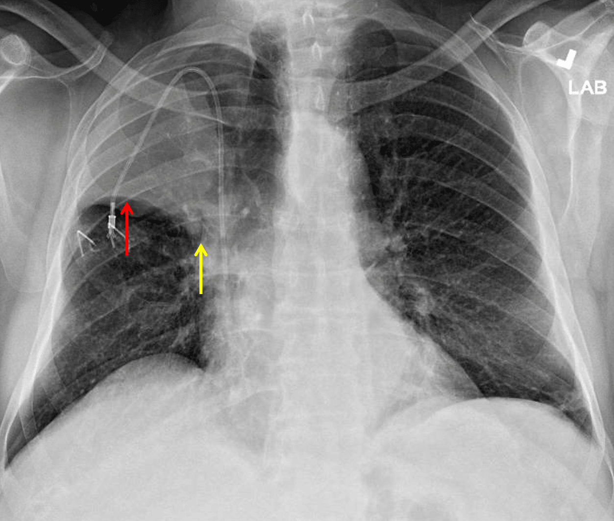
  • ์†Œ์—ฝ๊ฐ„ ์—ด๊ตฌ์˜ ์ƒ๋ฐฉ ์ „์œ„๋ฅผ ๋™๋ฐ˜ํ•œ ์šฐ์ƒ์—ฝ์˜ ํ˜ผํƒ ๋ฐ ์šฉ์  ๊ฐ์†Œ
  • ๊ฒฝ๋„์˜ ์šฐ์ธก ์ข…๊ฒฉ๋™ ํŽธ์œ„
  • ๊ฒฝ๋„์˜ ๋ฐ˜์ ์ƒ ์ขŒํ•˜์—ฝ ํ˜ผํƒ
  • ์ƒ๋Œ€์ •๋งฅ-์‹ฌ๋ฐฉ ์ ‘ํ•ฉ๋ถ€์— ์„ ๋‹จ์ด ๊ฑธ์น˜๋Š” ์šฐ์ธก ๋‚ด๊ฒฝ์ •๋งฅ ์ ‘๊ทผ Port-A-Cath
  • ํ‰๋ง‰ ์‚ผ์ถœ ๋˜๋Š” ๊ธฐํ‰ ์—†์Œ
  • ์‹ฌ๋‚ญ์‹ฌ์žฅ ์‹ค๋ฃจ์—ฃ์˜ ์ •์ƒ ํฌ๊ธฐ ๋ฐ ๊ตฌ์„ฑ

์ง„๋‹จ

์šฐ์ƒ์—ฝ ํ—ˆํƒˆ

์ƒ˜ํ”Œ ๋ณด๊ณ ์„œ

์šฐ์ƒ์—ฝ ํ—ˆํƒˆ๋กœ ์ ์•ก ๋งˆ๊ฐœ ๋˜๋Š” ์ค‘์‹ฌ๋ถ€ ํ์‡„์„ฑ ์ข…๊ดด๊ฐ€ ์šฐ๋ ค๋ฉ๋‹ˆ๋‹ค. ์ถ”๊ฐ€ ํ‰๊ฐ€๋ฅผ ์œ„ํ•ด ํ‰๋ถ€ CT๋ฅผ ๊ถŒ๊ณ ํ•ฉ๋‹ˆ๋‹ค.

์ขŒํ•˜์—ฝ์˜ ๊ฒฝ๋„ ๋ฐ˜์ ์ƒ ์Œ์˜์€ ๋น„ํŠน์ด์ ์ด๋ฉฐ ๋ฌด๊ธฐํ, ํก์ธ ๋˜๋Š” ํ๋ ด๊ณผ ๊ด€๋ จ๋  ์ˆ˜ ์žˆ์Šต๋‹ˆ๋‹ค.

๊ณ ์ฐฐ

  • ์šฐ์ƒ์—ฝ ํ—ˆํƒˆ์€ ์ข…์ข… ์ •๋ฉด ์ดฌ์˜์—์„œ ์†Œ์—ฝ๊ฐ„ ์—ด๊ตฌ์˜ ์ƒ๋ฐฉ ํŽธ์œ„๋ฅผ ๋™๋ฐ˜ํ•œ ์šฉ์  ๊ฐ์†Œ ๋ฐ ํ˜ผํƒ์œผ๋กœ ์‹๋ณ„๋ฉ๋‹ˆ๋‹ค
  • Golden S ์ง•ํ›„๋Š” ํ๋ฌธ๋ถ€ ์ข…๊ดด์— ์†Œ์—ฝ๊ฐ„ ์—ด๊ตฌ์˜ ๋‚ด์ธก ๋ถ€๋ถ„์ด ๊ฑธ๋ฆฌ๊ณ  ์ค‘๊ฐ„ ๋ฐ ์™ธ์ธก ๋ถ€๋ถ„์ด ์ƒ์Šนํ•  ๋•Œ ๋‚˜ํƒ€๋‚˜๋ฏ€๋กœ, ์ด๋Ÿฌํ•œ ์–‘์ƒ์ด ๋ณด์ด๋ฉด ์ค‘์‹ฌ๋ถ€ ํ์‡„์„ฑ ์ข…๊ดด๋ฅผ ์ƒ๊ฐํ•˜์‹ญ์‹œ์˜ค

์ฃผ์„์ด ์žˆ๋Š” ์ด๋ฏธ์ง€ ๋ฐ ์ผ๋Ÿฌ์ŠคํŠธ

Right upper lobe collapse. The minor fissure is elevated laterally (red arrow) but not as elevated centrally (yellow arrow) raising concern for a perihilar mass.
Right upper lobe collapse. The minor fissure is elevated laterally (red arrow) but not as elevated centrally (yellow arrow) raising concern for a perihilar mass.

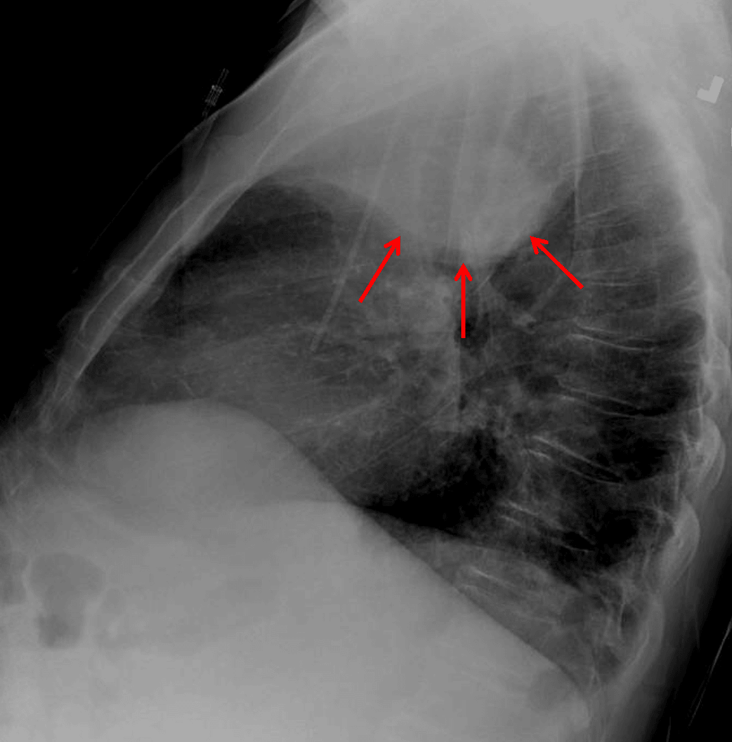
Right upper lobe collapse. Lateral view shows a paradoxical bulging appearance along the inferior margin of the collapsed lobe concerning for mass (red arrows).
Right upper lobe collapse. Lateral view shows a paradoxical bulging appearance along the inferior margin of the collapsed lobe concerning for mass (red arrows).

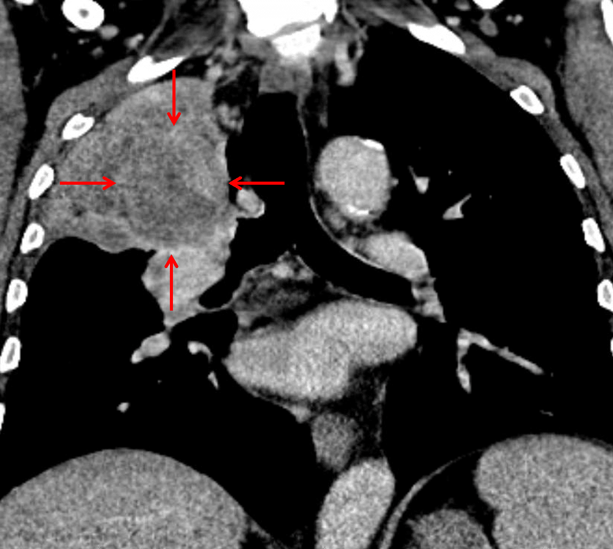
Subsequent CT confirmed the presence of a suprahilar mass with bronchial invasion (red arrows), though the mass is difficult to distinguish from the adjacent atelectatic lung.
Subsequent CT confirmed the presence of a suprahilar mass with bronchial invasion (red arrows), though the mass is difficult to distinguish from the adjacent atelectatic lung.

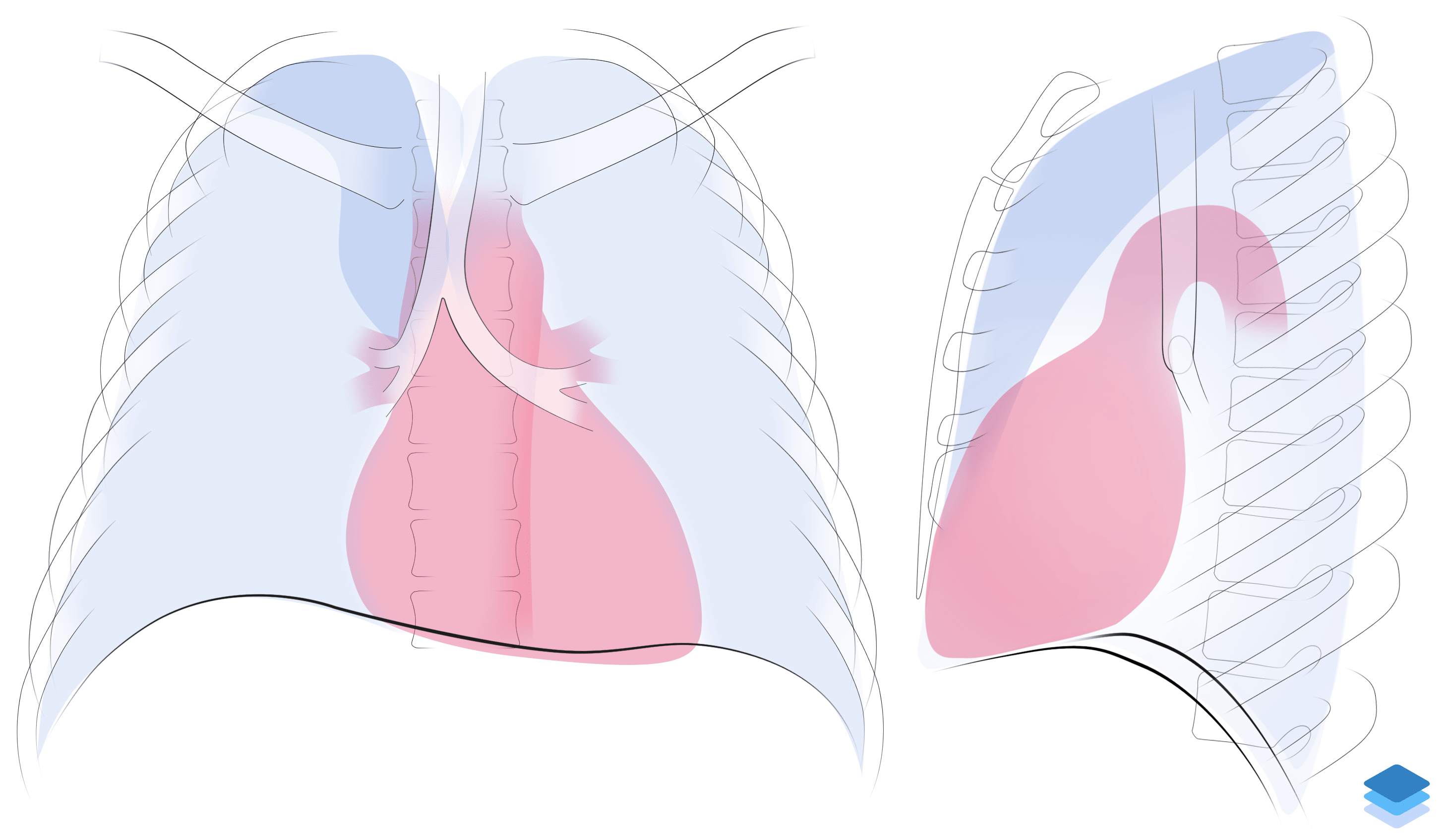
Right upper lobe collapse. Illustration by Valerie George, MD

Right upper lobe collapse. Illustration by Valerie George, MD

์ฐธ๊ณ ๋ฌธํ—Œ

์‚ฌ์ดํŠธ ํ”ผ๋“œ๋ฐฑ

์—ฌ๋Ÿฌ๋ถ„์˜ ํ”ผ๋“œ๋ฐฑ์€ CaseStacks ๊ฐœ์„ ์— ๋„์›€์ด ๋ฉ๋‹ˆ๋‹ค. ์˜๊ฒฌ, ์ œ์•ˆ ๋˜๋Š” ๋ฐœ์ƒํ•œ ๋ฌธ์ œ๋ฅผ ๊ณต์œ ํ•ด ์ฃผ์„ธ์š”.

/5
ํ”ผ๋“œ๋ฐฑ์„ ๋ณด๋‚ด์ฃผ์…”์„œ ๊ฐ์‚ฌํ•ฉ๋‹ˆ๋‹ค. ์†Œ์ค‘ํžˆ ๋ฐ˜์˜ํ•˜๊ฒ ์Šต๋‹ˆ๋‹ค.