マウスホイール、矢印キー、または左クリック(スクロールツール使用時)でスクロールしてください。
ui.case.use_touch_gestures
DICOMヘルプSource: Local (us-east1-c)
所見
- LEFT
- External ear:
- Mild microtia
- Atretic external auditory canal with a solid bony plate in the typical location of the external auditory canal measuring 5 mm in thickness
- Nonvisualization of the tympanic membrane
- Middle ear/mastoid:
- Mild asymmetric underpneumatization of the left mastoid temporal bone relative to the right
- Dysplastic handle of the malleus, which appears fixed to the anterior wall of the mesotympanum
- Normal appearance of the incus and stapes
- Normal volume and aeration of the middle ear
- Blunted appearance of the pyramidal eminence with a shallow facial recess
- Atypical splitting of the descending mastoid facial nerve canal with a larger channel exiting anteriorly
- Internal ear
- Normal appearance of the IAC, cochlea, and vestibular apparatus
- External ear:
- RIGHT
- Normal
診断
External auditory canal atresia
主要な画像所見
- External ear:
- Microtia commonly accompanies EAC atresia, but is well assessed clinically and less important for the radiologist to evaluate in detail
- The normal EAC is divided into cartilaginous and bony segments, should measure at least 4 mm in diameter, and usually has a mildly upslanting orientation from lateral to medial
- EAC atresia may be fibrous or bony – it is important to report the nature and width of the plate to aid with surgical planning
- Middle ear:
- The normal middle ear should measure at least 3 mm from cochlear promontory to the tympanic membrane or atretic plate
- A variety of ossicular anomalies can be seen, including abnormal morphology, abnormal articulation between the ossicles, and fixation to the walls of the middle ear or between ossicles. While the stapes is least frequently affected, an abnormal stapes is important to identify as this is the major factor in deciding between partial and total ossicular replacement protheses
- Although they are usually normal, it is important to assess the oval and round windows as these are necessary for successful
- An abnormal course of the facial nerve is common, particularly an inferior course of the tympanic segment overlying the oval window, placing the nerve at greater surgical risk. Abnormal branching of the mastoid segment can be seen, and in particular, an abnormally anterior course of the descending mastoid segment is important to note as this could be injured during canaloplasty
- There is a higher incidence of congenital cholesteatoma in these patients
鑑別診断
- None
臨床のポイント
- External and middle ear malformations often go together due to shared embryologic origins
- In cases of EAC atresia, try to determine if the atretic portion is bony or fibrous
- Make sure to assess for concomitant abnormalities of the ossicles, facial nerve course (particularly an inferior course of the tympanic segment and an anterior course of the mastoid segment), and abnormalities of the oval and round windows to aid your surgical colleagues
- Patients with EAC atresia are at increased risk for congenital cholesteatoma
考察
- External auditory canal (EAC) atresia exists on a spectrum of developmental anomalies encompassed in the broader term “congenital aural atresia”
- The first and second branchial arches (mesoderm) are responsible for formation of the middle and external ear, so EAC atresia is commonly accompanied by middle ear malformations
- In contrast, the inner ear develops from the otic placode (ectoderm) and is usually normal in cases of EAC atresia
- Although often an isolated finding, EAC atresia can occur with several syndromes, including:
- Crouzon syndrome (think of severe craniosynostosis and midface hypoplasia)
- Treacher Collins syndrome (think of mandibular and midface hypoplasia with retrognathia)
- Goldenhar syndrome (think of asymmetrically small hemifacial structures and vertebral segmentation anomalies)
- Pierre Robin syndrome (think of micrognathia, tongue retraction, and cleft or high arched palate)
- EAC atresia results in conductive, not sensorineural, hearing loss
- Management options include:
- Bone-anchored hearing aids to improve sound conduction to the inner ear
- Canaloplasty and tympanoplasty with or without ossicular chain reconstruction
注釈付き画像とイラスト
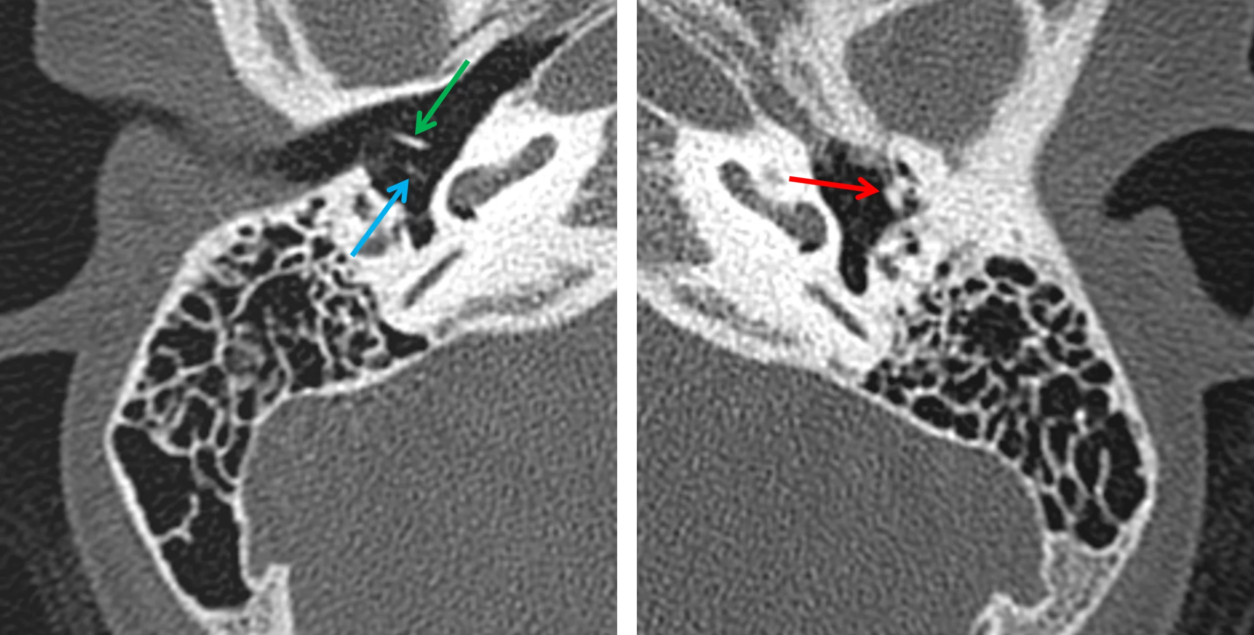
Normal morphology and orientation of the ossicles on the right (image on the left) with the handle of the malleus (green arrow) and long process of the incus (blue arrow) oriented perpendicular to each other. Abnormal morphology of the malleus and incus on the left (image on the right), which appear fused to the anterior wall of the middle ear (red arrow).
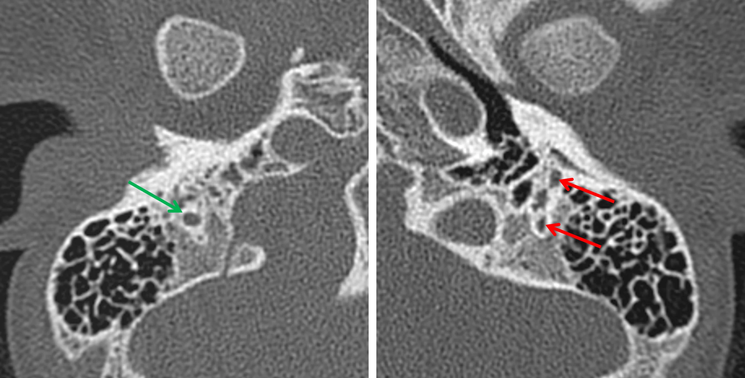
Normal course of the mastoid segment of the right facial nerve (image on the right, green arrow). Abnormal splitting of the mastoid segment of the left facial nerve with a larger canal coursing anteriorly (image on the right, red arrows).
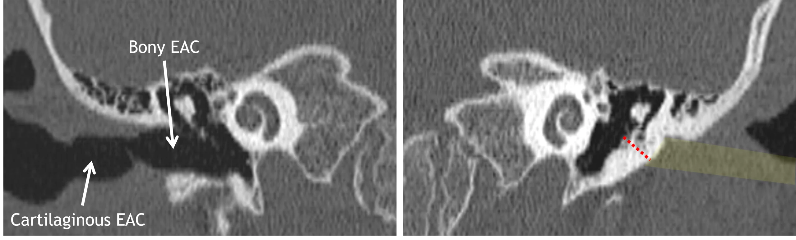
Normal right EAC (image to the left) with normal cartilaginous and bony segments. Atretic left EAC (image to the right), with the approximate expected location of the EAC highlighted in yellow and the red dotted line measuring the width of the bony plate where the bony portion of the EAC should be.
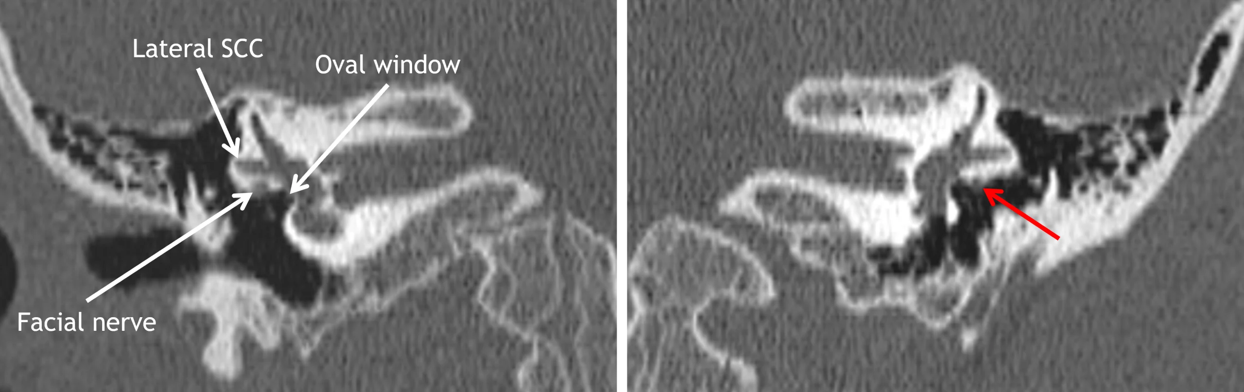
Normal right side (image on the left) with the tympanic segment of the facial nerve coursing immediately inferior to the lateral semicircular canal, not obstructing the oval window. Abnormal left side (image on the right) with an abnormally inferior, partially uncovered tympanic segment of the facial nerve (red arrow).
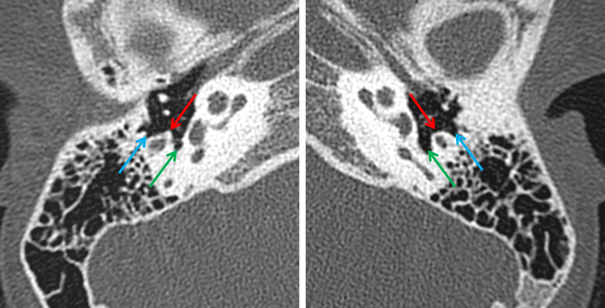
Normal right (image on the left and abnormal left (image on the right) middle ears. Blunted appearance of the pyramidal eminence (red arrows) and shallow appearance of the facial recess (blue arrows) on the left. Green arrows indicate the sinus tympani.