Use mouse wheel, arrow keys or left click (with scroll tool selected) to scroll
ui.case.use_touch_gestures
DICOM HelpSource: Local (us-east1-c)
Keyboard shortcuts (Alt+K)
Demographics:
44 years old, Female
Indication:
Knee pain
Findings
- No acute fracture
- Lateral patellar dislocation
- Moderate joint effusion
Diagnosis
Patellar dislocation
Sample Report
No acute fracture.
Lateral patellar dislocation.
Moderate joint effusion.
Joint spaces are maintained.
Discussion
- The patella most commonly dislocates laterally, which is often transient, such that there may be no radiographic abnormalities by the time the patient is imaged
- MRI often shows a “kissing contusion” pattern with bone contusions along the medial patella and lateral femoral condyle at the sites of impact
- MRI may also show associated injury to the medial patellofemoral ligament (MPFL) or medial retinaculum
- There are predisposing factors to patellar dislocation including trochlear dysplasia and patella alta, which will not be discussed further here
Annotated Images & Illustrations
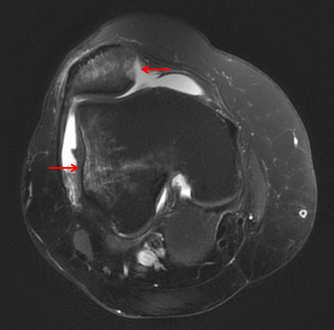
Sequela of lateral patellar dislocation. MRI in this patient shows kissing contusions in the medial patella and lateral femoral condyle (red arrows).