Age: 66
Sex: Female
Indication: Encephalopathy
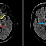
T2/FLAIR signal hyperintensity in the bilateral periaqueductal gray matter, medial thalami, hypothalamus, and possibly mamillary bodies. Though nonspecific, this distribution of signal abnormality raises particular concern for Wernicke encephalopathy. No evidence of acute ischemia.
Scattered T2/FLAIR hyperintensities in the periventricular and subcortical white matter, which though nonspecific are commonly attributable to chronic small vessel disease.
Extraaxial lesion along the lateral aspect of the right temporal lobe with low signal on all sequences, likely representing bulky dystrophic calcification. Consider postcontrast imaging to assess for associated enhancing mass.
250 cases
146 cases
372 cases
281 cases
84 cases
97 cases
43 cases
140 cases
75 cases
• Shuffle cases from our courses to simulate the mix of a call shift.
• Submit your own report before reviewing the case write-up.
250 cases
146 cases
372 cases
281 cases
75 cases
84 cases
97 cases
127 cases
43 cases
105 cases
13 cases
• Shuffle cases from our courses.
• Submit your own report.
• Earn up to 91 CME.
• Claim your CME to receive a certificate.
• Shuffle cases from our courses to simulate the mix of a call shift.
• Submit your own report before reviewing the case write-up.
• Earn up to 91 CME by completing cases in our radiology courses.
• Claim your CME to receive a certificate.
Reference database of normal imaging from birth to age 16
Summary of consensus guidelines for managing incidental CT findings
Index of select illustrations & videos from our courses
Visual reference for common mimics of pathology on CT
Reference database of normal imaging from birth to age 16
Summary of consensus guidelines for managing incidental CT findings
Index of select illustrations & videos from our courses
Visual reference for common mimics of pathology on CT
Pelvic MRI anatomy
Chest radiograph & CT anatomy
Abdominal CT anatomy
Cardiac CT anatomy
Brain & calvarial anatomy on CT/MRI
Cranial nerves on MRI
Shoulder MRI anatomy
Knee MRI anatomy
Resident/fellow-level anatomy
Pelvic MRI anatomy
Chest radiograph & CT anatomy
Abdominal CT anatomy
Cardiac CT anatomy
Brain & calvarial anatomy on CT/MRI
Cranial nerves on MRI
Shoulder MRI anatomy
Knee MRI anatomy
Resident/fellow-level anatomy
250 cases
146 cases
105 cases
13 cases
Coming soon
250 cases
146 cases
105 cases
13 cases
Coming soon
• Claim your CME to receive a certificate.
• Shuffle cases from our courses to simulate the mix of a call shift.
• Submit your own report before reviewing the case write-up.
• Earn up to 91 CME by completing cases in our radiology courses.
• Claim your CME to receive a certificate.
250 cases
146 cases
372 cases
281 cases
75 cases
84 cases
97 cases
140 cases
43 cases
• Shuffle cases from our courses to simulate the mix of a call shift.
• Submit your own report before reviewing the case write-up.
• Earn up to 91 CME by completing cases in our radiology courses.
• Claim your CME to receive a certificate.
View shortcuts |
Zoom/Pan |
Full screen |
Window/Level |
Expand/collapse |
Scroll |
Save the case |
Close case/tab |