Expand ("E")   Collapse
.

Age: 8 months

Sex: Female

Indication: Evaluate for tethered cord

Save ("V")

Lipomyelocele


Findings

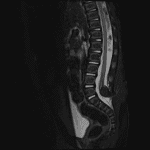
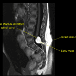
  • Low-lying spinal cord terminating at the level of L5
  • Associated T1/T2 hyperintense mass measuring 14 x 12 x 12 mm with signal suppression on STIR. This mass extends from the neural placode posteriorly through a spinal canal defect into the posterior paraspinal soft tissues
  • Sacral spinal dysraphism


Diagnosis

  • Lipomyelocele

Sample Report

Low-lying spinal cord terminating at the level of L5. Associated T1/T2 hyperintense mass measuring 14 x 12 x 12 mm with signal suppression on STIR, suggesting fatty composition. This mass extends from the neural placode posteriorly through a spinal canal defect into the posterior paraspinal soft tissues. These findings are consistent with a lipomyelocele.

Sacral spinal dysraphism.


Discussion

  • Neural tube defects can be complicated to understand and distinguish
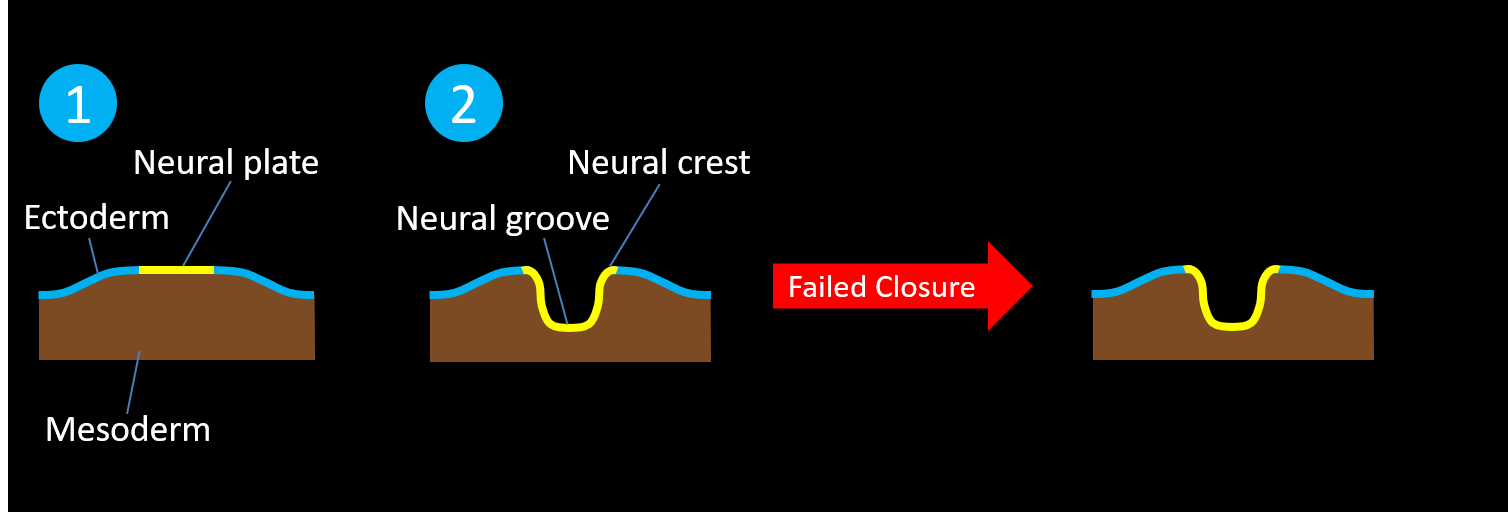
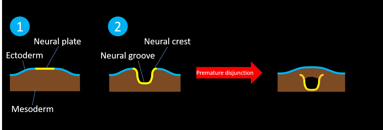
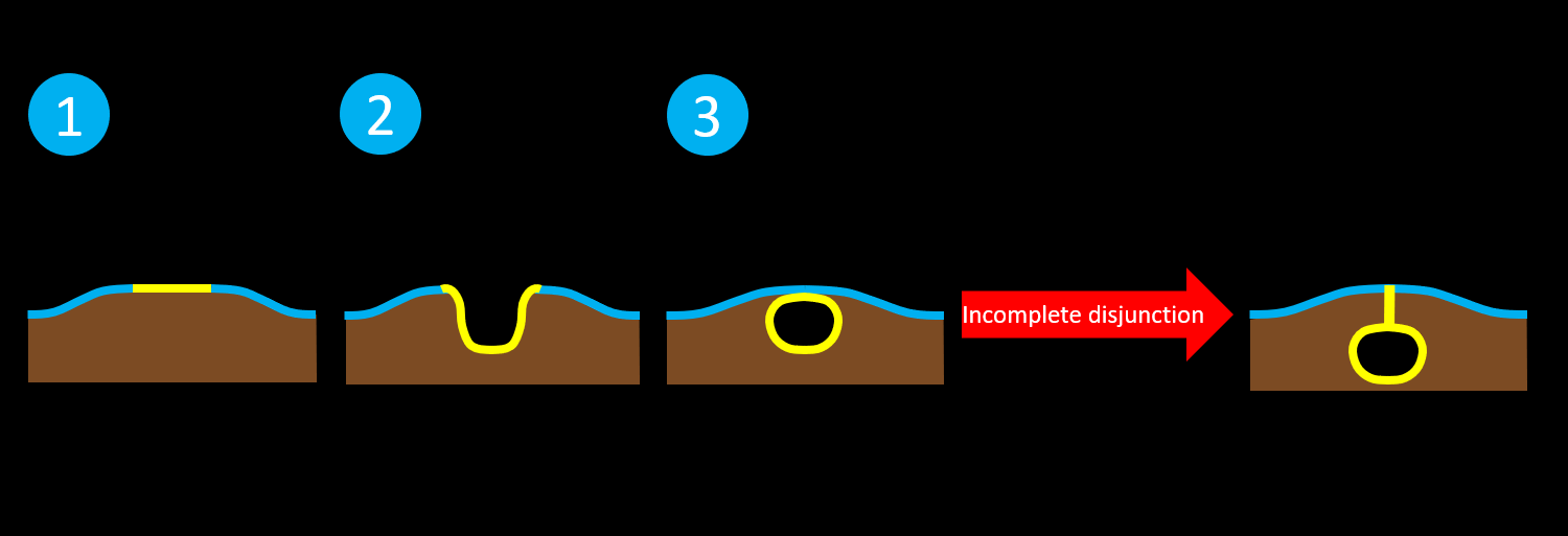
  • Try thinking about them in groups of conditions resulting from failures of different steps of neurulation (see below diagrams):
    • Normal neurulation occurs during the third and fourth weeks of gestation as the neural tube forms within and then separates from the ectoderm
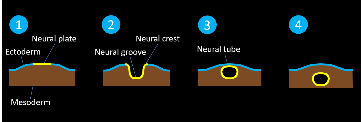
      • Failed closure and failed separation of the neural tube from the ectoderm results in open neural tube defects (i.e. defects with no skin covering), including:
        • Myelomeningocele – the meningeal lining and the neural placode extend beyond the skin surface
        • Myelocele – the neural placode remains flush with skin surface

      • Premature disjunction results in closed neural tube defects (i.e. defects with skin covering) where mesodermal elements are interposed within an incompletely closed neural tube that separates from the ectoderm, including:
        • Lipomyelomeningocele – lipoma-placode interface located outside of the spinal canal
        • Lipomyelocele – lipoma-placode interface located inside of the spinal canal
        • Meningocele – meningeal lining herniates through a spinal canal defect without herniation of neural elements
        • Terminal myelocystocele – syringocele herniates into a meningocele

      • Incomplete disjunction results in a closed neural tube defect with a persistent dermal sinus attached to the neural tube
        • May have associated intradural dermoid or epidermoid lesions

      • Additional disorders of abnormally located mesodermal elements can result in intradural lipomas without associated dural defects
  • This case is an example of a lipomyelocele because there is an intact skin covering, a fatty mass extending through a spinal canal defect, and the lipoma-placode interface is located within the spinal canal

Images

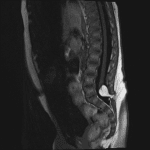
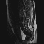
Low-lying spinal cord with associated fatty mass extending through a posterior defect in the spinal canal. The neural placode-mass interface is located within the spinal canal, consistent with a lipomyelocele. Low-lying spinal cord with associated fatty mass extending through a posterior defect in the spinal canal. The neural placode-mass interface is located within the spinal canal, consistent with a lipomyelocele. Low-lying spinal cord with associated fatty mass extending through a posterior defect in the spinal canal. The neural placode-mass interface is located within the spinal canal, consistent with a lipomyelocele. This case is a lipomyelocele because there is an intact skin covering, a fatty mass extending through the posterior elements, and the lipoma-placode interface is located within the spinal canal.


📣 Feedback?

⌨️  Keyboard Shortcuts ("K")

Call Preparation

Prepare for call efficiently with interactive cases, sample reports, and annotated images.

1,400+ cases  91.25 CME

Call Simulator

   Neuro CT

250 cases

  Neuro MRI

146 cases

   Peds Radiographs

372 cases

   MSK Radiographs

281 cases

   Chest CT

84 cases

   Chest Radiographs

97 cases

   KUB

43 cases

   Body CT

140 cases

   Ultrasound

75 cases

Call Simulator

• Shuffle cases from our courses to simulate the mix of a call shift.
• Submit your own report before reviewing the case write-up.

Neuro Fellowship

Reviews of neuro topics with clinical pearls, differentials, and in-depth discussions.

131 cases  13.1 CME

   Brain Tumors

105 cases

   Neurodegenerative

13 cases

   Congenital Hearing Loss

13 cases

Call Preparation

   Neuro CT

250 cases

   Neuro MRI

146 cases

   Peds Radiographs

372 cases

   MSK Radiographs

281 cases

   Ultrasound

75 cases

   Chest CT

84 cases

   Chest Radiographs

97 cases

   Body CT

127 cases

   KUB

43 cases

Neuro Fellowship

   Brain Tumors

105 cases

   Neurodegenerative

13 cases

Call Simulator

• Shuffle cases from our courses.
• Submit your own report.

CME

• Earn up to 91 CME.
• Claim your CME to receive a certificate.

Call Simulator

• Shuffle cases from our courses to simulate the mix of a call shift.
• Submit your own report before reviewing the case write-up.

CME

• Earn up to 91 CME by completing cases in our radiology courses.
• Claim your CME to receive a certificate.

On Call

Quick references created specifically to help while on call.

  Peds Normals by Age

Reference database of normal imaging from birth to age 16

   Incidental Findings

Summary of consensus guidelines for managing incidental CT findings

   Media Index

Index of select illustrations & videos from our courses

  Neuro CT Mimics

Visual reference for common mimics of pathology on CT

On Call

  Peds Normals by Age

Reference database of normal imaging from birth to age 16

   Incidental Findings

Summary of consensus guidelines for managing incidental CT findings

   Media Index

Index of select illustrations & videos from our courses

  Neuro CT Mimics

Visual reference for common mimics of pathology on CT

Anatomy

Labelled radiographs and CT/MRI series teaching anatomy with a level of detail appropriate for medical students and junior residents.

   Pelvis

Pelvic MRI anatomy

   Chest

Chest radiograph & CT anatomy

   Body

Abdominal CT anatomy

   Cardiac

Cardiac CT anatomy

   Brain

Brain & calvarial anatomy on CT/MRI

   Cranial Nerves

Cranial nerves on MRI

   Shoulder

Shoulder MRI anatomy

   Knee

Knee MRI anatomy

   Temporal Bone

Resident/fellow-level anatomy

Anatomy

   Pelvis

Pelvic MRI anatomy

   Chest

Chest radiograph & CT anatomy

   Body

Abdominal CT anatomy

   Cardiac

Cardiac CT anatomy

   Brain

Brain & calvarial anatomy on CT/MRI

   Cranial Nerves

Cranial nerves on MRI

   Shoulder

Shoulder MRI anatomy

   Knee

Knee MRI anatomy

   Temporal Bone

Resident/fellow-level anatomy

Neuro Courses

Includes our call preparation neuro courses (Neuro CT and Neuro MRI) and our neuro fellowship courses.

   Neuro CT

250 cases

  Neuro MRI

146 cases

   Brain Tumors

105 cases

   Neurodegenerative

13 cases

   Congenital Hearing Loss

Coming soon

Neuro Courses

   Neuro CT

250 cases

   Neuro MRI

146 cases

   Brain Tumors

105 cases

   Neurodegenerative

13 cases

  Congenital Hearing Loss

Coming soon

CME

• Claim your CME to receive a certificate.

Call Simulator

• Shuffle cases from our courses to simulate the mix of a call shift.
• Submit your own report before reviewing the case write-up.

CME

• Earn up to 91 CME by completing cases in our radiology courses.
• Claim your CME to receive a certificate.

   Neuro CT

250 cases

   Neuro MRI

146 cases

   Peds Radiographs

372 cases

   MSK Radiographs

281 cases

   Ultrasound

75 cases

   Chest CT

84 cases

   Chest Radiographs

97 cases

   Body CT

140 cases

   KUB

43 cases

Call Simulator

• Shuffle cases from our courses to simulate the mix of a call shift.
• Submit your own report before reviewing the case write-up.

CME

• Earn up to 91 CME by completing cases in our radiology courses.
• Claim your CME to receive a certificate.

⌨️ Keyboard Shortcuts

View shortcuts

Zoom/Pan

  Full screen

  Window/Level

  Expand/collapse

  Scroll

  Save the case

  Close case/tab

Previous Slice (PC)

Next Slice (PC)

SHIFT Previous Slice (MAC)

 SHIFT Next Slice (MAC)

 Toggle presets

 Previous series (if multiple)

 Next series (if multiple)

  Previous case (⌘ ◄ on a mac)

  Next case (⌘ ► on a mac)

Toggle dark mode