Age: 54
Sex: Male
Indication: Left nasal mass
Save ("V")
Inverted Papilloma
Diagnosis
- Inverted papilloma
 Sample Report
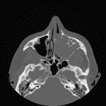
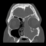
Expansile mass in the left maxillary sinus with associated sinus wall remodeling and dehiscence and bulging into the nasal cavity and retroantral fat with obstruction of the left ostiomeatal unit. Hyperostosis about the left infraorbital canal is typical for inverted papilloma and represents the likely site of tumor origin.
Expansion of the medial aspect of the left frontal sinus with polypoid soft tissue markedly thinning the intersinus septum and bulging into the right frontal sinus, which could represent contiguous extension of the maxillary sinus mass versus separate mass/mucocele.
No definite dehiscence through the inner table of the calvarium.
Discussion
- When assessing opacified sinuses, look for evidence of a mass including bowing, thinning, or destruction of the sinus walls
- Inverted papillomas often have associated hyperostotic pedicles, which may be sessile or conical in morphology
- One retrospective review of 76 surgically-confirmed cases of inverted papilloma found associated focal hyperostosis on CT in 63%, with areas of focal hyperostosis corresponding with the tumor origin/attachment site in 89%
- Although inverted papillomas can grow very large, the attachment sites are usually small, potentially allowing less aggressive surgery if the attachment site can be identified preoperatively
- Inverted papillomas require surgical removal for symptomatic reasons as well as for the 10% rate of synchronous or metachronous squamous cell carcinoma


 View shortcuts
 Zoom/Pan
 Full screen
 Window/Level
 Expand/collapse
 Scroll
 Save the case
 Close case/tab


 Previous series (if multiple)
 Next series (if multiple)
