Age: 66
Sex: Female
Indication: Trauma
Save ("V")
Case #8
Findings
Chest radiograph
- Widening of the superior mediastinum
- Hazy opacification of the left hemithorax, which may relate to airspace opacification and/or layering pleural fluid
CT
- Chest
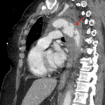
- Complex acute aortic injury just distal to the origin of the left subclavian artery with external contour irregularity and large surrounding mediastinal hematoma
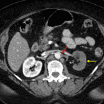
- Additional site of acute aortic injury just below the diaphragmatic hiatus with distal extension into and occlusion versus near occlusion of the left renal artery. No large surrounding hematoma at this location
- Mediastinal hematoma displaces and mildly narrows the trachea, inferiorly displaces the left mainstem bronchus, and extends around the esophagus in the posterior mediastinum
- Moderate-sized left hemothorax
- Small right pleural effusion
- Dependent groundglass opacities in both lungs
- Abdomen/Pelvis
- Decreased enhancement of the interpolar region and lower pole of the left kidney
- Heterogeneous enhancement of the right kidney
- Mild fat stranding adjacent to the left common iliac vein
- Flattened appearance of the IVC
- Focal fatty infiltration in the liver adjacent to the falciform ligament
- Cholecystectomy changes with mild intrahepatic and extrahepatic biliary duct dilation
- Small volume free fluid layering in the anatomic pelvis
- Hysterectomy
- MSK
- Acute nondisplaced fracture of the left L1 transverse process
Diagnosis
- Acute traumatic aortic injury
 Sample Report
Severe acute traumatic aortic injury just distal to the origin of the left subclavian artery with external contour irregularity concerning for full-thickness mural tear. Limited evaluation for active bleeding as no delayed images were obtained through the chest, but there is a large surrounding mediastinal hematoma and moderate-sized left hemothorax.
Additional site of aortic injury just below the diaphragmatic hiatus with extension into and occlusion versus near occlusion of the left renal artery. No large surrounding hematoma. Resultant ischemia of the majority of the left kidney.
Heterogeneous enhancement of the right kidney is likely the result of platelet aggregation in the setting of traumatic aortic injury.
Probable injury of the left common iliac vein.
Flattening of the IVC concerning for hypovolemia.
Acute nondisplaced left L1 transverse process fracture.
Discussion
📣 Feedback?
⌨️ Keyboard Shortcuts ("K")
Help | Terms | Privacy Policy | Cookie Policy
Medical Disclaimer | © 2024 CaseStacks LLC
Related Cases
References
- Heneghan RE, Aarabi S, Quiroga E, Gunn ML, Singh N, Starnes BW. Call for a new classification system and treatment strategy in blunt aortic injury. J Vasc Surg 2016; 64(1): 171-176.
- Kaewlai R, Avery LL, Asrani AV, Novelline RA. Multidetector CT of blunt thoracic trauma. Radiographics 2008; 28: 1555-1570.
- Steenburg S, Ravenel J, Ikonomidis J, Schonholz C, Reeves S. Acute traumatic aortic injury: Imaging evaluation and management. Radiology 2008; 248: 748–762.


 View shortcuts
 Zoom/Pan
 Full screen
 Window/Level
 Expand/collapse
 Scroll
 Save the case
 Close case/tab


 Previous series (if multiple)
 Next series (if multiple)
