Age: 65
Sex: Male
Indication: Trauma
Save ("V")
Case #4
Findings
- Chest
- Anterior mediastinal hematoma, measuring up to 1.5 cm in thickness
- Anterior right upper lobe and lateral right middle lobe pulmonary contusions
- Mild bilateral dependent groundglass opacities
- Trace right hemopneumothorax
- Abdomen/Pelvis
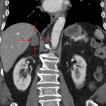
- High density 3.2 x 2.2 cm right adrenal lesion
- Gallbladder distension without radiopaque gallstones, gallbladder wall thickening, or pericholecystic inflammatory changes
- Mild intrahepatic and extrahepatic biliary duct dilation
- Diffuse fatty atrophy of the pancreas with several punctate parenchymal calcifications
- 1.3 cm left renal cyst
- MSK
- Acute manubrial fracture with 3 mm cortical offset
- Acute fractures of the right first through eighth ribs including displaced fractures of the first, sixth, and seventh ribs
- Acute nondisplaced/minimally displaced fractures of the left second through eighth ribs
- Acute nondisplaced fractures of the right superior and inferior pubic rami and left inferior pubic ramus/ischium. The right superior pubic ramus fracture extends into the pubic root and right hip joint
- No diastasis of the pubic symphysis or sacroiliac joints
- Right flank soft tissue contusion with a small area of internal hyperdensity which enlarges on delayed phase images
- Subcutaneous gas and hematoma adjacent to the right first rib fracture in proximity to the subclavian vessels without evidence of active hemorrhage at this location
Diagnosis
- Adrenal hematoma
 Sample Report
Acute mildly displaced manubrial fracture with a subjacent mediastinal hematoma measuring 1.5 cm in thickness. No direct evidence of acute aortic injury.
Right first through eighth and left second through eighth rib fractures with a trace right hemopneumothorax. No left pneumothorax.
Right upper and middle lobe contusions. Bilateral dependent groundglass opacities likely represent a combination of aspiration and atelectasis.
High density right adrenal lesion likely representing a hematoma. Recommend followup adrenal protocol abdomen CT in 6-8 weeks to ensure resolution.
Bilateral obturator ring injuries without evidence of unstable pelvic trauma. The right superior pubic ramus fracture extends into the hip joint, which remains located.
Right flank soft tissue contusion with evidence of active hemorrhage. This area should be amenable to direct pressure.
Nonspecific mild biliary dilation. Recommend correlation with LFTs.
Sequela of chronic pancreatitis.


 View shortcuts
 Zoom/Pan
 Full screen
 Window/Level
 Expand/collapse
 Scroll
 Save the case
 Close case/tab


 Previous series (if multiple)
 Next series (if multiple)
