Age: 73
Sex: Male
Indication: Left flank pain
Save ("V")
Case #50
Findings
- Lower chest
- Circumferential mural thickening of the lower thoracic esophagus
- Moderate centrilobular and paraseptal emphysema
- Mild bilateral subpleural reticulation
- Abdomen/Pelvis
- 13 mm proximal left ureteral calculus with associated severe left hydroureteronephrosis and severe renal cortical thinning
- Multiloculated fluid and gas collection in the left perinephric space with posterior rupture/extension into the posterior pararenal space and left flank soft tissues measuring 15 x 13 x 30 cm
- Subcentimeter hypodensity in the right hepatic lobe which is too small to characterize
- Splenomegaly
- Mild circumferential mural thickening of the second portion of the duodenum
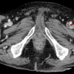
- Nonocclusive filling defect in the left femoral vein
- Possible additional nonocclusive filling defect in the right femoral vein
- Atherosclerotic calcification of the abdominal aorta and branch vessels without aneurysm
- MSK
- L2 vertebral body compression fracture with near complete central height loss but no bony retropulsion
- Multilevel degenerative changes of the spine, most advanced at L4-L5 and L5-S1
Diagnosis
- Perinephric abscess
- Deep venous thrombosis
 Sample Report
Obstructing 13 mm proximal left ureteral calculus with resultant severe left hydronephrosis, severe left renal cortical thinning, and a large multiloculated left perinephric abscess which extends into the left flank soft tissues. A nuclear medicine renogram could be considered to assess differential renal function if this will impact the decision for surgical management.
Nonocclusive left, and possibly right, femoral deep venous thrombosis.
Age-indeterminate L2 compression fracture with near complete central height loss but no bony retropulsion.
Nonspecific mural thickening of the second portion of the duodenum, which could represent a focal duodenitis in the correct clinical setting.
Circumferential thickening of the lower thoracic esophagus, which can be seen with reflux esophagitis.


 View shortcuts
 Zoom/Pan
 Full screen
 Window/Level
 Expand/collapse
 Scroll
 Save the case
 Close case/tab


 Previous series (if multiple)
 Next series (if multiple)
